PHD Veterinary Service

PHD Veterinary Service

Contact Info

Dr. Porter @ 352-258-3571
portermi.dvm@gmail.com

Read more about Dr. Porter
And PHD Veterinary Services @


Showing posts with label chronic cough. Show all posts

Friday, July 27, 2012

Equine Pharyngitis!!


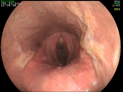
The image above was from a 10 year old gelding that presented for a history of coughing and mild nasal discharge. The cough was intermittent and would vary from day to day. Recently, the cough had worsened as had the summer heat! Based on the history, the most likely cause was predicted to be heaves (AKA: COPD). However, when the endoscope was introduced into the gelding's upper airway it was clear that we were dealing with something quite different. There was evidence of pharyngeal scarring (cicatrix) and active pharyngeal ulceration  in a 360 degree (red circle below) range.  These findings were consistent with active inflammation and chronic changes. In addition, there was chronic, active inflammation of the arytenoids (green arrow).


The cause of pharyngeal inflammation/ulceration varies and is often never determined. In some cases, it may be complicated by bacterial infection. In some cases, the inciting cause is presumed to be an environmental irritant that the horse is chronically exposed to. The chronic inflammation results in scarring or cicatrix formation that can result in narrowing of the pharynx. Pharyngeal cicatrix formation is a serious complication that has no reliable remedy!  As such, a chronic couch (>2 weeks) should be evaluated via endoscopy prior to the development of scarring. Pharyngeal inflammation is typically treated with forced rest and topical antibiotics/steroids in the form of a nasal flush. 


Friday, July 13, 2012

Cleft Palate!

Image 1
The first two images are from a yearling thoroughbred that presented for a long history of nasal discharge, coughing while eating, and the presence of water exiting from nostrils while drinking. The filly had been treated with antibiotics prior to the exam. Endoscopic exam revealed a defect within the soft palate that extended nearly the entire length of the soft palate. In addition, the epiglottis was entrapped by the aryepigltottic fold. 

Image 2
 The defect within the soft palate is known as a "cleft palate" and most likely has been there since birth. Fortunately, this is a relative rare condition! Normally, the palate plays the role of separating the nasal passages from the oral passage. Hence, a horse is an obligate nasal breather and the palate keeps water and feed from entering the airway. When a cleft palate exists, there is communication between the air passage and oral passage resulting in feed and water going the "wrong way". This may consist of feed material exiting the nostril and/or traveling down the trachea into the lungs. If these horse go undiagnosed they are typically "poor doers" with failure to thrive secondary to chronic respiratory infections. Sadly, there is little to be done to help such a severe cleft palate. Less severe cases may be addressed with surgical intervention with mixed results. 


Image 3

The third image if that of a "normal" horse. The epiglottis is visible and is resting on top of the complete soft palate. Diagnosis of a cleft palate is often made immediately after birth during a thorough post-foaling exam. The veterinarian should palpate the length of the hard and soft palate with their finger to make sure it is normal. If missed at foaling, the diagnosis can be made quite simply with an endoscopic exam.