PHD Veterinary Service

PHD Veterinary Service

Contact Info

Dr. Porter @ 352-258-3571
portermi.dvm@gmail.com

Read more about Dr. Porter
And PHD Veterinary Services @


Friday, August 30, 2013

Pleuropneumonia in a Horse

A 17 year-old gelding QH presented for a 3 week history of intermittent fever and coughing.

History:

The gelding had been treated with 3 different antibiotics over the past 3 weeks and there were no improvements in clinical signs. The problem started after the horse had been transported approximately 12 hours via truck and trailer. Within 24 hours of arriving to the show grounds the gelding was sick!

Physical exam:

When I examined the horse, the gelding was in distress! His heart rate was 80 bpm, respiratory rate was 60-70 bpm (shallow) and his body temperature was 102 degrees. Auscultation of the thorax noted lung sounds (sound of air moving in and out of lungs) on both sides of the horse only ABOVE the level of the shoulder but lung sounds were absent or muffled below the level of the shoulder. Suspecting pleuropneumonia, I performed a trans-thoracic ultrasound exam. A significant amount of fluid was noted in the pleural space along with large fibrin tags and multiple abscesses! ( Figure 1-3).

     
Figure 1

 In the video clips below, the fibrin tags can be seen "floating" in the excessive fluid within the pleural space. In addition, the fluid appears as "cellular" suggesting a heavy component of fibrin and purulent debris (pus).


In Figure 2-3, large abscesses are noted adjacent to the body wall. Ultimately, these abscess would need to be exteriorized through a rib resection in order for the horse to completely heal. Unfortunately, the ultrasound findings combined with the severe physical distress were very poor prognostic indicators and the owner elected humane euthanasia!  There was near zero chance that this horse could have been saved regardless of the medical and surgical intervention provided!
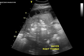
Figure 2


Figure 3
Pleuropneumonia in also called pleurisy and refers to bacterial infection of the pleural cavity and the surrounding soft tissue structures. This condition is VERY deadly and originates from bacterial colonization of the lower airway! The condition is more common in young horses that are transported long distances on a regular basis. Hence the term "shipping fever". A common belief is that horses which are transported with their head tied and a bag of hay in front of them are predisposed because they are not able to properly clear their airway of airborne debris and pathogens. Normally, horses eat with their head down which minimizes the passage of unwanted matter down their trachea and into their lungs.  This condition can be treated effectively if diagnosed early, i.e, within 1-2 days of clinical signs! Aggressive medical treatment is a must and includes IV antibiotics, chest drainage/lavage, and supportive care. One of the most painful horses I witnessed as a resident was a young filly with pleurisy!! Looked like a bad case of colic but was in fact pleuropneumonia!!


Friday, August 23, 2013

Aortic Valve Insufficiency in a Horse

Figure 1

Figure 2A and 2B
 The image in Figure 1 is of a horse's heart taken via trans-thoracic ultrasound. Specifically, the ultrasound probe is centered on the aortic valve. The horse presented for a prepurchase exam and a loud murmur was detected during the physical exam. There was no history of exercise intolerance nor was there evidence of heart failure during the physical exam. The murmur was best described as diastolic (during the filling phase) and included a high musical pitch at the end of the murmur. During the cardiac ultrasound exam, color flow doppler was imaged (Figure 1) to determine the flow of blood across the aortic valve. Normally, the color of the blood flow will be blue or red depending on whether the blood is moving towards or away from the ultrasound probe. When there is a "leaky" valve or a valve that is "insufficient" the color of the blood flow is bright yellow, white or orange. This color flow pattern is consistent with turbulence across the valve due to blood traveling "backwards" through the valve. In Figure 2, the red arrow identifies the normal direction of blood flow through the aortic valve. In Figure 3 a small white line is highlighted by the yellow arrows. This white line corresponds to one of the 3 valves that make up the aortic valve. In this horse, the aortic valve is thickened resulting in a "leaky" valve. The "leakiness" can be noted in Figure 4 as bright yellow and white blood flow in the opposite direction of normal blood flow across the aortic valve. The thickened valve likely vibrates as the jet of blood flows "backwards" causing the musical heart murmur!  Aortic insufficiency is the most common type of heart murmurs in horses and is often benign during the early phases of development. However, if the horse lives long enough, the aortic insufficiency can lead to heart failure in the horse. As such, heart murmurs in horses should not be dismissed as "normal" and inconsequential but should be carefully documented via ultrasound exam!!


Figure 3


Figure 4


Friday, August 16, 2013

Equine Metabolic Syndrome

The images in Figures 1 and 2 are of an obese donkey. If you follow the dark, dorsal stripe you will notice that the withers of this donkey completely falls to the left side due to the large concenetration of fat that has accumulated within this area!! This condition is also termed "cresty neck".  In Figure 2, the abnormal fat accumulation over the donkey's top line is apparent as is the large deposit of fat just in front of the tail head region! Although slightly humurous, this condition presents a serious medical problem for this donkey. The abnormal accumulation of fat in the area of the neck, top-line, tail head and internally can predispose donkeys and horses to a condition known as metabolic syndrome.

Figure 1

Figure 2
Metabolic syndrome occurs most commonly in over-conditioned horses but has a species predilection for Morgans horses, Arabian horses, Tennessee walking horses, and ponies. The condition is some what similar to adult onset diabetes in humans, which also results in a state of insulin resistance. The serious and some times deadly complication of metabolic syndrome is laminitis!! (Figure 3 and 4). The association between insulin resistance and laminitis is not well understood however in humans with adult onset diabetes, the high circulating levels of glucose are toxic to blood vessels and may cause loss of limb due to poor perfusion. This has not been proven in horses but sounds very similar to what happens in the case of laminitis!  These cases typically present to me AFTER the horse has already foundered and there is a long history (6-24 months) of intermittent lameness, coffin bone rotation, and recurrent foot abscesses!! In my experience, these horses will NOT improve until the insulin resistance is documented and treated!! Unfortunately, if the condition is not diagnosed early enough, severe coffin bone rotation may occur resulting in protrusion of the coffin bone through the bottom of the foot and humane euthanasia (Figure 3 and 4) !


Figure 3


Diagnosis simply involves a blood sample collected from a fasted animal that compares the level of glucose and insulin simultaneously! Occasionally, a glucose challenge may be necessary to establish that a horse is glucose intolerant and insulin resistant. Once diagnosed, the treatment involves regulating the caloric intake of the animal and medical management with thyroid hormone supplement. It is very important to understand that these horses do NOT have a thyroid deficiency!! We are merely increasing the metabolic demand by treating with thyroid hormone supplementation and forcing the animal to lose body fat!!  In addition, to the medical management, the caloric intake is carefully regulated between limited pasture access and low starch diets. Finally, the farrier plays a integral part in helping those horses that have foundered due to the metabolic syndrome. Interestingly, these horses often remain tender-footed regardless of the type of shoe UNTIL the insulin resistance is properly regulated. 

You can read more about equine metabolic syndrome at:


Friday, August 9, 2013

Chronic Obstructive Airway Disease in a Horse

The endoscopic image in Figure 1 is from within a horse's trachea. In Figure 1, the endoscope is positioned just in front of the main bifurcation of the trachea where it splits into the right and left bronchi (Figure 2) . This is just in front of the horse's heart also known as the carina! The purpose of passing an endoscope down to this level of the horse's respiratory tract is to evaluate for the accumulation of debris (feed material, pus and/or blood) and to perform a diagnostic procedure called a bronchoalveolar lavage (BAL). In this particular case, the horse presented for a chronic cough and exercise intolerance.


Figure 1

Figure 2

As the endoscope was passed down the trachea there was a moderate amount of white foamy fluid along the ventral aspect of the trachea (Figure 2 and 3). This debris is consistent with sputum that originates within the lung tissue and is coughed into the trachea. This finding can be consistent with either pneumonia (bacterial infection) or an allergic airway condition that is also known as COPD/Heaves! To determine the cause of the sputum, a BAL is performed. In this procedure, the endoscope is passed into the primary bronchi until it is lodged within the bronchi. At this point a small volume of sterile saline is passed into the bronchi through the scope and then collected via aspiration. The fluid is analyzed to determine the percentage of different cell types and from this data, it can be determined if the horse has an infection or allergic airway disease.

Figure 2

Figure 3

The importance of determining which disease process is causing the sputum is that treatment for allergic airway disease involves systemic corticosteroid administration which will lower the horse's immune system and significantly worsen any bacterial infection the horse may have!! Hence, it is key to perform the BAL  prior to any treatment to determine if the horse needs antibiotics or steroids or maybe antibiotics plus steroids!! The BAL can be performed stall-side with a sedated horse.