PHD Veterinary Service

PHD Veterinary Service

Contact Info

Dr. Porter @ 352-258-3571
portermi.dvm@gmail.com

Read more about Dr. Porter
And PHD Veterinary Services @


Showing posts with label mobile equine veterinarian. Show all posts

Friday, February 27, 2015

Subcutaneous Pythiosis in a Horse

.A 10 year-old warmblood gelding presented to PHD veterinary services for the complaint of a swollen left front fetlock that was associated with a chronic draining wound. The recent history included a biopsy of the wound and diagnosis of pythiosis per histopathology. The gelding was treated with a single dose of a systemic pythiosis vaccine. In Figures 1 and 2 the yellow arrows are pointing to the firm swelling that is directly on the backside (palmar aspect) of the fetlock joint. In addition, the red arrow in Figure 2 corresponds to the chronic draining wound. The gelding was sensitive to direct pressure over the swelling, passive flexion of the fetlock and was mildly lame at the trot.
.
.
Figure 1

Figure 2
An ultrasound exam was performed of the area in question. The firm swelling consisted of multiple fluid filled structures along with focal areas of apparent mineralization. The structure appeared embedded within the distal annular ligament; however the superficial flexor tendon (SDF) and the deep digital flexor tendon (DDF) appeared to be normal. In Figure 3 the yellow arrows are highlighting the fluid filled structures. This case is a very unusual presentation for pythiosis in a horse. The majority of cases present with an external lesion as seen in Figure 4. As external lesions there are several treatment options which involve aggressive surgical de-bulking and various topical medications. The pathogen responsible for this condition is found in stagnant water and invades superficial abrasions that might be present any where on the horse. Hence the lower limbs are most commonly affected but it has been diagnosed in the naso-pharynx and GI track of horses.

Figure 3


Figure4
Because of the complicated location of the lesion in this horse, we elected to treat with a potent anti-fungal medication known as Amphotericin B. The medication was administered through a vein in the lower limb using regional limb perfusion. This technique maximizes the local perfusion of the soft tissues with the medication and minimizes the systemic effect of the medication. 

Figure 5
The lesion was re-evaluated 30 days after the initial treatment and there was approximately 40-50% reduction in size of the lesion. However, after 60 days, the lesion appeared to resume growth and a second treatment with Amphotericin B was recently administered. In addition, the gelding as received 2 vaccines against the pythiosis and will likely receive another. Because of the location, surgery is not an option for this horse hence the systemic therapy is our best option at this point. The case is on -going and the blog will be updated as we go!!


Figure 6

Saturday, January 17, 2015

Eye Lid Tumor in a Donkey

A 2 year-old female donkey presented to PHD Veterinary services for the complaint of multiple tumors surrounding the left and right eye. The tumors have been present for several months and have been treated by surgical debulking and intra-lesional injections with a variety of chemotherapy agents. However, the tumors continue to return. There is a golf ball size tumor just below the right eye and the left eye is nearly closed due to the tumor's involvement of the entire eye lid!
.
.

The plan for this donkey involves biopsy of the tumors followed by local treatment with an immune stimulant that contains mycobaterial cell wall extract. PHD veterinary services will be working with Dr. Brendan Mangan from Affiliated Veterinary Specialist in Gainesville, Florida. Dr. Mangan is a board certified ophthalmologist with extensive experience treating such cases! Dr. Mangan suspects that these tumors are sarcoids and not squamous cell carcinomas. Certainly, an unusual presentation for sarcoids in a young donkey!! Dr. Mangan submitted several core biopsies this week for confirmation and began treatment with the mycobaterial cell wall extract.


After the initial treatment with the immune stimulant, Dr. Mangan plans to surgically remove the tumors surrounding both eyes! Thereafter, it is likely that the injection of the local immune stimulant will be repeated along with focal cryotherapy. The biopsy results and follow-up pics will be added to this post next week so stay tuned!!!


Friday, November 14, 2014

Hock Arthritis in a Horse

A 14 year-old gelding presented to PHD veterinary services for the complaint of left hind limb lameness. The lameness had been noted for 6 months with minimal clinical improvement after several months of pasture rest. On presentation the gelding was a grade 3/5 lame in the left hind limb when trotted in a straight line. Flexion of the lower limb and upper limb did NOT worsen the lameness. In addition, the lameness did NOT worsen when lunged in either direction. Physical exam of the left hind limb did not identify any swelling or joint effusion however the gelding's range of motion of the hock joint was reduced. Radiographic evaluation of the limb was elected as the best option for identifying the source of lameness in this horse.  In Figure 1 and 2, the lower hock joints are identified as the following: PIT= proximal intertarsal joint, DIT= distal intertarsal joint, and TMT= tarsal metatarsal joint. 

Figure 1

Figure 2

In Figure 3, the lower hock joints are circled in blue and the yellow arrows are highlighting areas of significant arthritis across the front of the lower hock joints. For comparison, in Figure 4, the right hock is imaged in a radiograph and there is no evidence of arthritis in any of the hock joints.

Figure3
Figure 4

In Figure 4, the front of the lower hock joints appear disfigured (yellow arrows) by the advanced arthritis present. In addition, there is evidence of arthritis in the most proximal hock joint (tibio-tarsal joint) which significantly worsens the prognosis for this horse (Figure 4, red arrow).


Figure 4
The cause of such advanced arthritis in only one hock suggests trauma or a previous infection. Unfortunately, the full history of this horse was not available. Recommendations included a daily anti-inflammatory medication and intra-articular therapy with corticosteroids. Prognosis is guarded for soundness due to the involvement of the tibio-tarsal joint and the degree of arthritis noted in the lower hock joints.

Friday, October 10, 2014

Fractured Tail Bone in a Horse

A seven year-old gelding presented for a history of recent trauma via a pasture mate over the region of the tail head. The gelding was able to move his tail however there was firm swelling around the region and he was tender to palpation (Figure 1 and 2). Physical exam performed 2 weeks after the injury was first noted revealed moderate swelling yet no pain on palpation. In addition, the tail had a normal range of motion and there was normal tail anal tone.
.
Figure 1

Figure 2

A radiographic exam was performed and a mildly displaced fracture of the first coccygeal bone was identified. In Figure 3, the vertebrae in the center of the image has what appears to be a cap on the spinous process which represents the mildly displaced fracture. In Figure 4, the coccygeal vertebrae in question is magnified and the fractured bone is highlighted with the red arrows. The yellow arrow in Figure 4 identifies the fracture line through the spinous process of the first coccygeal vertebrae.

Figure 3

Figure 4
The fracture will heal with time and there should not be any negative, long term effects from this injury. However, there remains a concern of the development of a sequestrum (de-vitalized bone) at the fracture site. A sequestrum would develop if the fracture resulted in loss of blood supply to the fractured fragment. This would result in the likely development of a draining fistula as the body attempts to reject the sequestrum. The gelding will be given several weeks of rest and relaxation before returning to light riding. A follow-up radiographic exam will be performed in 2-4 months.

Friday, September 26, 2014

Fungal Plaque in a Horse

A 7 year-old gelding presented to PHD veterinary services for the complaint of mild epistaxis (bloody nasal discharge). Endoscopic exam of the nasal passages identified a golden colored mass (Figure 1) that was covered in blood and was located within the opening to the ethmoid turbinates. The mass was diagnosed as an ethmoid hematoma based on location, appearance, and behavior. The client was given the option of surgical removal or treatment with intra-lesional doses of formalin. Based on the relatively small size of the ethmoid hematoma it was decided to attempt treating 1-2x with formalin and if there was not complete resolution then surgical resection would be pursued.


Figure 1
Four weeks after the first injection of formalin, a follow-up endoscopy noted significant reduction in the size of the ethmoid hematoma (Figure 2) and what appeared to be a second mass deeper within the ethmoid turbinates. The second ethmoid hematoma was not visualized during the initial exam because the first ethmoid hematoma was blocking the view! Based on these findings, the second ethmoid hematoma was injected with formalin in a similar fashion as the original ethmoid hematoma.

Figure 2
Six weeks after the second formalin injection the client reported that there was a slight yet persistent bloody discharge from the affected nares. Endoscopic exam revealed what appears to be a fungal plaque (black/white/yellow) adhered to the site of the ethmoid hematoma (Figure 3 and 4). This is an unusual finding and may prove to be a challenging complication. Fungal plaques have a predilection for vascular tissue and can infect the upper airway of horses. Fungal infection within the guttural pouch of a horse is well documented and can result in a catastrophic hemorrhage if not diagnosed and treated early. Fungal infection within the ethmoid turbinates is not common in my experience. Often these fungal plaques do NOT respond to systemic anti-fungal medication and must be either removed or treated aggressively with topical medication.
.
Figure 3

Figure 4
The current clinical plan for this horse is medication with oral anti-fungal medication for several weeks. If there is no change, the fungal plaques will be treated with topical anti-fungal medication. Stay tuned....

Finally after 4 weeks of anti-fungal medication, there is no evidence of a fungal plaque and there is NO evidence of an ethmoid hematoma (Figure 5). The ethmoid turbinates are visible for the first time in this horse since the initial exam. Although there is no evidence of an existing ethmoid hematoma, these tumors commonly reoccur hence the gelding will be monitored closely for the next 12 months. 


Figure 5

Friday, September 19, 2014

Coffin bone rotation and Sinking in a donkey.

Sadly, I share the story of my very own donkey named "Lollipop". She is approximately 12 years old and every summer gets a bit foot sore when the green grass is lush. Through benign neglect and some luck, she has recovered every year with just a few days of phenylbutazone treatment! This year has been different. She has been lame in left front foot for over 2 months. Initially, she was lame in both front feet and I kept her feet supported with impression material and bandages. After 30 days of foot bandages, I switched to Soft Ride boots with removable orthotics designed for acutely foundered horses. The donkey remained lame in the left front foot hence I broke down and radiographed both front feet. On the day that the radiographs were taken, I noted that the entire coronary band was soft and painful in the left front foot!!
.
.
Figure 1

Figure 2
.Figure 1 is a radiographs of the left front foot and Figure 2 is of the right front foot. There is evidence of chronic laminitis in both front feet with coffin bone rotation and pedal bone lysis. However, the most concerning observation was the radiographic evidence of coffin bone "sinking", predominantly in the left front foot. This occurs when the lamina becomes detached all the way around the foot and can result in the entire hoof sloughing off the foot!! In Figure 3 and 4, the red line corresponds to the coronary band and the blue arrow is the distance from the coronary band to the coffin joint. In Figure 3 (left front foot), the blue arrow is longer than the blue arrow in Figure 4. This would suggest that the entire bony column has "sunk" into the hoof capsule. This would also explain why the coronary band was soft and painful. In essence, Lollipop is trying to lose her hoof!  If this was to happen she would likely require humane euthanasia!!

Figure 3
 
Figure 4
Due to the severity of her condition, she is now locked up in a stall and I have placed her left front foot in a cast. The cast will hopefully stabilize the foot/hoof and allow time for healing of the lamina. She is comfortable on 1/2 gram of phenylbutazone 2x per day and other than being annoyed about the stall confinement, she appears stable. Stay tuned and keep her in your thoughts and prayers!!

Saturday, August 16, 2014

Drug Testing and Prepurchase Exams for Horses

A 10 year old, warm-blood mare presented to PHD veterinary services for a prepurchase exam. The standard exam, complete with limb flexion was performed and the mare was found to be completely sound and negative to limb flexion. The buyer requested baseline radiographs of both hocks and both front feet. In addition, it was strongly recommended that a drug screen of the mare's blood be performed for the detection of sedatives, anti-inflammatory medications and corticosteroids.
.
.
.
Figure 1

Although the mare was sound and did not resent limb flexion, there was radiographic evidence of advanced osteoarthritis of the lower hock joints.  In Figures 1,2, and 4, the yellow arrows are pointing to radiographic changes consistent with arthritis along the front or dorsal aspect of the lower hock joints. There is new bone growth (osteophytes) and joint space narrowing. In addition, in Figure 3, the blue arrows are pointing to the area of sclerosis surrounding the lower hock joints which suggests chronicity of the arthritis and the apparent "fusion" of the distal hock joints. These radiographic changes are advanced and are surprising considering that the mare was sound for the prepurchase exam.

Figure 2

Figure 3
Even though the mare was sound, I did NOT pass this horse for sale and intended use based on radiographic findings and advised the buyer to at least wait for the drug screen results prior to making their decision!   The standard drug screen takes approximately 5-7 days for results to be reported. Interestingly, the mare's blood tested positive for high doses of an anti-inflammatory medication which likely explains why this horse was sound and negative to limb flexion even though she has advanced arthritis in the lower hock joints. This case represents yet another example of the importance of drug testing and base line radiographs for prepurchase exams!! Admittedly, it is a financial slippery slope once you begin the radiographic exam regarding how many areas to evaluate, however; areas of high probablity such as the hocks and front feet should always be considered!

Figure 4

Saturday, August 9, 2014

Allergic Airway Disease in horses.

In the past 30 days (July 8 - August 8) I have examined 10 horses for the complaint of coughing and poor performance. The horses have ranged in age from 8 to 19 years of age. There has been no commonality with regards to sex or breed. However, they all live in Florida and it is the hottest month of the year!  Through a series of diagnostics which include a re-breathing exam, upper airway endoscopy, and trans-tracheal wash, all 10 horses have been diagnosed with allergic airway disease (AKA: heaves or COPD). None of these horses had previously been diagnosed with this condition. 
.
.
Upper trachea with large amounts of sputum present.

Lower trachea with thickened mucosa due to chronic inflammation

Allergic airway disease in horses typically results in coughing, increased respiratory effort, increased "abdominal" breathing, exercise intolerance and weight loss. If not managed properly, the condition worsens from year to year and can result in the death of the horse!! I have posted a blog previously discussing this disease and how it should be diagnosed and treated. Please click on the link below to read my blog from last year regarding this condition.

Allergic Airway Disease in a Horse


Friday, June 20, 2014

Summer Sore Time is here!!

Although I have already written a blog concerning summer sores in horses (go to: http://michaelporterdvm.blogspot.com/2013/06/habronemiasis-summer-sore-in-horse.html) , the condition is a recurring one and it deserves another visit!! Below are several images from different horses that suffer from recurring habronemiasis or equine summer sores. The horse in Figure 1 suffers from recurring habronemiasis in all 4 limbs, whereas the horse in Figure 2 recently developed summer sores along his sheath!! These summers sores are medium in size and treatable with topical and systemic medication. However the horse in Figure 3 is suffering from a horrible summer sore of his lower limb that has not been treated but allowed to grow unabated and permanently disfigure the limb. It is frustrating to the horse owner that these lesions tend to return every year with the return of flies however if treated early and aggressively, management of this condition is quite do-able!!

Figure 1

Figure 2
For horses with recurring or first time habronemiasis, I recommend the following:

1: Systemic treatment with a dewormer containing Ivermectin. Treat twice, approximately 3-4 weeks appart.
2: Topical treatment with Dr. Porter's summer sore cream!!
3: If the summer sore is excessively large or in a location that is difficult to treat with topical medication, I recommend systemic therapy with corticosteroids. I prefer to medicate with a tapering dose of oral prednisolone over 30 days.

It is critical that the barn management incorporate a fly control program of their choosing. In addition, horses which suffer from recurring summer sores need to be treated the moment there is any redness or mild irritation present in the old summer sore sites!! If treated early, the lesion will typically respond favorably to topical medication without the need for systemic corticosteroids! 

Figure 3

Friday, June 13, 2014

Fractured Spinous Processes in a Horse

A 10 year-old gelding arrived to a horse show and was seriously injured during the unloading from the trailer. The rear gate was lowered and the horse remained standing in the trailer with the "butt bar" in place (Figure 1). For some unknown reason, the horse panicked and attempted to unload with the butt bar in place. The horse became trapped below the butt bar and struggled for several minutes before the owner was able to release the bar. Within moments of the event, the horse's withers became swollen and very sensitive to the touch. The gelding was treated with systemic anti-inflammatory agents and returned home in the trailer WITHOUT the  butt bar!


Figure 1
Two weeks after the injury, the gelding presented to PHD veterinary services for a radiographic evaluation of the withers. In Figure 2, the green line outlines the withers of the horse. The bone-like fingers projecting upward below the green line are the spinous processes which make up the withers. On physical exam there was minimal swelling over the withers however the horse was very sensitive to any pressure and there appeared to be a "dip" in the very bottom of the withers (yellow arrows).

Figure 2
Radiographic evaluation of the "dip" in the withers confirmed multiple fractures of 3 spinous processes (yellow arrows in Figure 3). The first spinous process that is fractured appears to be in several fragments and displaced from its normal position. The second spinous process appears fractured but not displaced and the third process appears to have an avulsion of the most proximal aspect of the spinous process. Although it is likely that these fractures will heal in time and be some what stable, the current concern is the development of a frustrating condition known as fistulated withers. This  occurs when a piece of fractured bone becomes devitalized of blood supply and subsequently "dies" becoming a sequestrum. The body's natural response is to rid itself of the sequestrum and will develop a draining wound that originates from the sequestrum. These wounds do NOT resolve until the sequestrum has been removed and this can be extremely challenging from a surgical stand point. Currently there is not evidence of a sequestrum however it will take several weeks/months to determine if one will develop. Prognosis for return to riding will depend the development of fistulated withers and how comfortable the horse is once the fractures have stabilized.

Figure 3

Friday, May 23, 2014

Severe Cartilage Degeneration in a Horse

A 12 year-old show jumper mare presented to PHD veterinary services for the history of recurring forelimb lameness. In the past 2 years, the mare had the left front coffin joint treated with corticosteroids and hyaluronic acid every 6 months. However, the most recent treatment with intra-articular corticosteroids did not result in resolution of the lameness. On presentation, the mare was a grade 3/5 lame in the left front. The lameness was most evident when trotting on firm ground and in a circle to the right. The mare was NOT positive to hoof tester exam and was mildly positive to lower limb flexion. The coffin joint was "blocked" with carbocain and the mare's lameness improved by >85%. Radiographic exam of the left front foot only revealed evidence of mild osteoarthritis of the coffin joint.  (Figure 1).
Figure 1
Due to the following factors: 1) a history of poor response to intra-articular therapy, 2) a positive response to blocking the coffin joint, and 3) minimal data via the radiographic exam, it was recommended that the distal forelimb of this mare be evaluated via magnetic resonance imaging (MRI).  The MRI exam requires general anesthesia and generally costs between $1800-$2000; however the information gathered from an MRI exam is far superior than any other type of diagnostic exam! As such, I strongly recommend considering an MRI exam in cases that the CAUSE for the lameness has not been determined and a reasonable attempt has been made to treat and/or diagnose the problem. Figures 2-4 includes several images from the MRI exam involving this show jumper mare. A small, blue circle is included in each image to high light the large articular cartilage and subchondral bone defect that was discovered through the MRI study. In each image, the lesion appears as a small black circle at the surface of the bone. In addition to this finding, the MRI study noted additional evidence of chronic degeneration of the coffin joint. The full extent of the degeneration and the cartilage damage was NOT evident in the radiographic exam!!

Figure 2

Figure 3

Figure 4
Although the prognosis was poor for return to soundness, the mare was treated aggressively with various regenerative therapies and extended rest. Unfortunately, the mare remained significantly lame and it was determined that humane euthanasia was the best option due to the relentless foot pain. A post-mortom exam was performed and a very large area of abnormal cartilage loss (blue circle in Figure 5) was noted that corresponded to the MRI findings. This case represents yet another exam of the importance for getting an answer as to WHAT is causing the problem such that a prognosis can be provided followed by a proper course of action.

Figure 5
In the state of Florida, the most sensitive MRI for imaging in horses is ONLY available at the University of Florida's College of Veterinary Medicine. In addition, the board certified radiologists at the Veterinary College of Veterinary Medicine are especially well trained for the evaluation of MRI studies in horses! For more information about their services please go to:

http://largeanimal.vethospitals.ufl.edu/services/diagnostic-imaging/


Friday, May 9, 2014

Superficial Flexor Tendonitis in a Horse


A 12 year old mare presented for sudden onset forelimb lameness and swelling of the tendons in the right front limb. On palpation, the swollen tendons were significantly sensitive to palpation yet it was not possible to determine if it was the superficial flexor tendon (SDF) or the deep digital flexor (DDF) tendon. The mare was a grade 3/5 lame in the right front when trotted in a straight line on firm ground. Ultrasound exam of the soft tissue structures revealed a core-like lesion within the outside corner of the SDF tendon. In Figures 1-3, the core-like lesion appears as a black area (red circle) within the body of the SDF tendon (blue circle).  In Figure 2, the image to the right is a cross-sectional view of the tendon and the lesion is a small black circle within the body of the SDF tendon. In the image to the left in Figure 2, the tendon is in a longitudinal view and the lesion appears as a black streak through the body of the SDF tendon.This view is important because it highlights the over-all length of the lesion which in this case was 4-5cm long. The cross-sectional area measured in Figure 3 determined that the core-like lesion was approximately 20% of the entire cross-sectional area of the SDF tendon.

Figure 1

Figure 2

The mare was treated with ultrasound guided injection of Platelet rich plasma (PRP), stall rest, daily ice therapy and we will follow-up with 3 doses of extra-corporeal shock wave treatment. We anticipate a full return to work after the appropriate period of rehabilitation.

Friday, May 2, 2014

On-Line Pharmacy for PHD Veterinary Services!!


The above medications are just a few of the hundreds that my clients will be able to purchase via my on-line pharmacy!! The pharmacy will be open for business within the next 2 weeks and can be accessed at the following address:

PHD Veterinary Services On-Line Pharmacy

Once open for business, my clients will be able to register their pet's information and order the medications desired. I will receive an instant email to confirm authorization for the sale of the medications and the client will enter their credit card information. This pharmacy will contain hundreds of medications and nutraceuticals for cats, dogs, and horses! Once ordered the products will ship direct to my clients for quick delivery.

I anticipate that PHD Veterinary Service's on-line pharmacy will offer my clients an efficient and affordable method to purchase prescribed medications for their cats, dogs, and horses. There will be monthly sales on some of the more popular products so please visit my blog site to find out when the pharmacy is open for business!! Please visit the on-line pharmacy today and register your cats, dogs, and horses! 

Friday, March 28, 2014

Ethmoid Hematoma versus Paranasal Sinus Cyst in a Horse


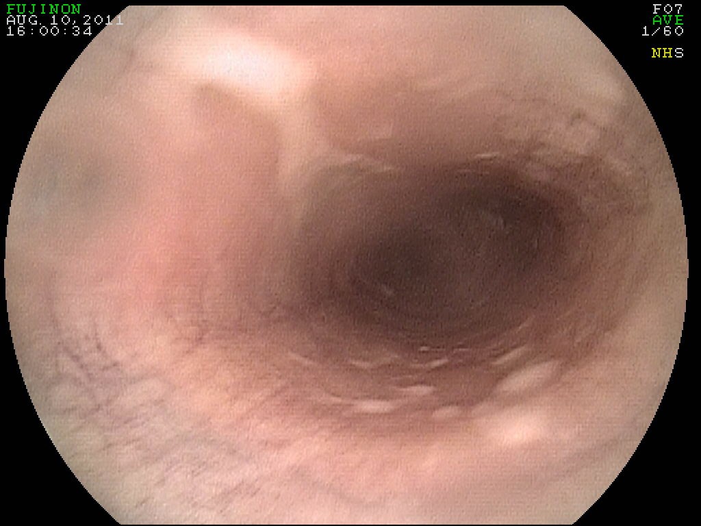
Two horses presented to PHD Veterinary services for the same complaint of "no air moving through one of the horse's nasal passages!" Both horses had a history of mild to moderate nasal discharge that had increased slowly over the past 6 months. On presentation, a simple evaluation of air passing through the nasal cavities revealed that there was NO air moving through the affected side on each horse however, endoscopic exam revealed a unique problem in each horse.

Figure 1
 In Figure 1, a smooth, white soft tissue mass was identified within 2 inches of the opening of the nasal cavity. This soft tissue mass was completely obstructing the nasal passage. In addition, when the scope was passed through the unaffected side, the soft tissue mass was noted to be extending into the naso-pharynx suggesting that the soft tissue mass extended through out the entire nasal passage (Figure 2). In Figure 2, the white wall of tissue noted along the right side of the image is the soft tissue mass as it extends into the naso-pharynx.The soft tissue mass is most likely consistent with a paranasal sinus cyst however surgical removal will be required to confirm the diagnosis. These types of cysts develop in young horses and grow slowly over months and years until a clinical problem develops. Surgical removal provides complete resolution of these types of cysts!!

Figure 2


Figure 3
In the second horse, a large golden-colored soft tissue mass was identified in the region of the nasal passage closest to the naso-pharynx. There was more discharge associated with this soft tissue mass and small areas of hemorrhage were noted. The soft tissue mass was completely obstructing the nasal passage and was originating from the ethmoid turbinate region which most likely classified it as a ethmoid hematoma!! These types of tumors typically present with a complaint of a unilateral bloody nasal discharge for months before they completely obstruct the nasal passage. However, they can be fast growing tumors and require an aggressive approach to eradicate. Treatment may involve either surgical removal or repeated injections of formalin. It has been my experience, having injected several horses for YEARS, that the tumors tend to return with this approach! Hence, I recommend surgical removal when first diagnosed, especially if the tumor is invading the sinus cavity or the naso-pharynx.