PHD Veterinary Service

PHD Veterinary Service

Contact Info

Dr. Porter @ 352-258-3571
portermi.dvm@gmail.com

Read more about Dr. Porter
And PHD Veterinary Services @


Showing posts with label COPD. Show all posts

Saturday, August 9, 2014

Allergic Airway Disease in horses.

In the past 30 days (July 8 - August 8) I have examined 10 horses for the complaint of coughing and poor performance. The horses have ranged in age from 8 to 19 years of age. There has been no commonality with regards to sex or breed. However, they all live in Florida and it is the hottest month of the year!  Through a series of diagnostics which include a re-breathing exam, upper airway endoscopy, and trans-tracheal wash, all 10 horses have been diagnosed with allergic airway disease (AKA: heaves or COPD). None of these horses had previously been diagnosed with this condition. 
.
.
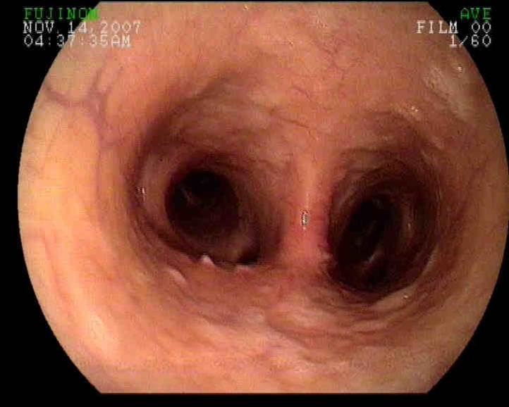
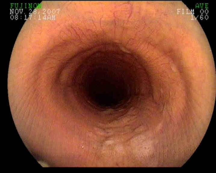
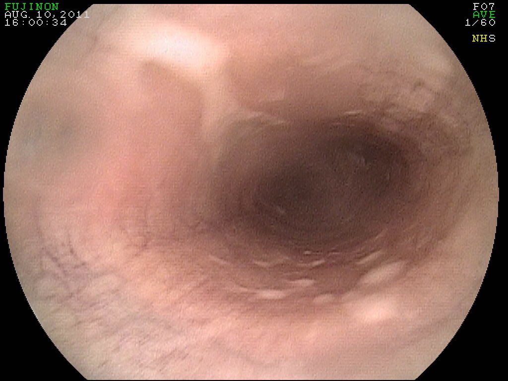
Upper trachea with large amounts of sputum present.

Lower trachea with thickened mucosa due to chronic inflammation

Allergic airway disease in horses typically results in coughing, increased respiratory effort, increased "abdominal" breathing, exercise intolerance and weight loss. If not managed properly, the condition worsens from year to year and can result in the death of the horse!! I have posted a blog previously discussing this disease and how it should be diagnosed and treated. Please click on the link below to read my blog from last year regarding this condition.

Allergic Airway Disease in a Horse


Friday, October 11, 2013

Respiratory Disease in a Horse

A 12 year-old mare presented for a history of increased respiratory rate and effort during the late summer months. In addition, a white discharge was noted from both nares. At presentation the mare had labored breathing, significantly nasal flaring and there was a large abdominal component to her respiratory effort (see videos below). Auscultation of the lungs noted wheezes and crackles and a very mild cough was elicited when the larynx was palpated. The client was of limited financial means and asked that I perform the fewest tests necessary to determine the source of the problem! As such, we opted to begin with an upper airway endoscopy to determine if there was any abnormality within the larynx and if not take a look down the upper trachea. The larynx was normal however there was a significant amount of white debris within the upper trachea (Figures 1-2). This white debris is otherwise known as sputum and is produced within the lungs by inflammatory cells!


Figure 1

Figure 2
The sputum present within the trachea may be due to an allergic airway condition known as heaves/COPD or it may be due to broncho-pneumonia!  These conditions present with similar symptoms but are treated quite differently. Hence making the correct diagnosis is essential! There are several diagnostic tests which will evaluate the lungs and the fluid within the lungs however the one exam that will give you all the information necessary to determine the cause of the sputum and how to treat is a trans-tracheal wash.  The trans-tracheal wash is performed by catheterizing the trachea through a small incision that is made along the ventral aspect of the neck, approximately half way between the horse's throat latch and thoracic inlet. The catheter is passed into the trachea and down to the level of the lungs. A small volume of sterile saline is flushed into the lungs and then aspirated out for analysis. This sample of fluid should contain the sputum noted in the endoscopy exam. The sputum is analyzed for cell type and for the presence of bacteria. If bacteria are found then the sample is tested for which antibiotic is most ideal to treat the pneumonia. This procedure is easily performed in the field with mild sedation and proper technique!


In this case, the mare was noted to be suffering from heaves AND pneumonia. This is not common but does occur and requires very specific medication to save the horse's life!! The horse was treated with a combination of systemic corticosteroids and appropriate antibiotics. Clinically, the horse presented as a typical heaves case however if we had only treated with corticosteroids the broncho-pneumonia would have worsened significantly and potentially caused the death of the patient!!

Friday, August 9, 2013

Chronic Obstructive Airway Disease in a Horse

The endoscopic image in Figure 1 is from within a horse's trachea. In Figure 1, the endoscope is positioned just in front of the main bifurcation of the trachea where it splits into the right and left bronchi (Figure 2) . This is just in front of the horse's heart also known as the carina! The purpose of passing an endoscope down to this level of the horse's respiratory tract is to evaluate for the accumulation of debris (feed material, pus and/or blood) and to perform a diagnostic procedure called a bronchoalveolar lavage (BAL). In this particular case, the horse presented for a chronic cough and exercise intolerance.


Figure 1

Figure 2

As the endoscope was passed down the trachea there was a moderate amount of white foamy fluid along the ventral aspect of the trachea (Figure 2 and 3). This debris is consistent with sputum that originates within the lung tissue and is coughed into the trachea. This finding can be consistent with either pneumonia (bacterial infection) or an allergic airway condition that is also known as COPD/Heaves! To determine the cause of the sputum, a BAL is performed. In this procedure, the endoscope is passed into the primary bronchi until it is lodged within the bronchi. At this point a small volume of sterile saline is passed into the bronchi through the scope and then collected via aspiration. The fluid is analyzed to determine the percentage of different cell types and from this data, it can be determined if the horse has an infection or allergic airway disease.

Figure 2

Figure 3

The importance of determining which disease process is causing the sputum is that treatment for allergic airway disease involves systemic corticosteroid administration which will lower the horse's immune system and significantly worsen any bacterial infection the horse may have!! Hence, it is key to perform the BAL  prior to any treatment to determine if the horse needs antibiotics or steroids or maybe antibiotics plus steroids!! The BAL can be performed stall-side with a sedated horse.

Friday, May 3, 2013

Arytenoid Chondritis in a Horse


A 12 year old quarter horse mare presented for a 2-3 month history of coughing and "wheezing". The mare had developed the symptoms some what suddenly and was responsive to systemic corticosteroids. As such, it was initially thought that the mare was developing signs of heaves or COPD (chronic obstructive pulmonary disease). Heaves is relatively common in the southeastern United States and is a chronic progressive condition that involves an allergic condition that affects the lungs and causes fibrosis and scarring.     At presentation, the client was interested in finding out for sure what was causing the coughing and wheezing. Re-breathing exam was essentially normal with no wheezing detected or coughing. The mare was currently being treated with corticosteroids and had been for the past 48 hours. The initial plan was to scope the mare and perform a bronchoalveolar lavage (BAL) to confirm the diagnosis of heaves. However, when the endoscope was passed to the point of the larynx, we discovered what was the true problem!


Figure 1
In Figure 1 and 2, the larynx of the horse in question appears grossly abnormal. In Figure 1, the horse is in between breaths and there is thin slit between the arytenoids which consists of her airway. In Figure 2, the mare is taking a deep breath and the full extend of her airway is probably the diameter of a sharpie pen! There is large linear ulcer that crosses the left arytenoid cartilage and the significant thickening of both left and right arytenoid cartilages suggest chronic inflammation. In Figure 3, a "normal" endoscopy demonstrates what the full extent of the airway should be during inspiration and what normal arytenoid cartilage should look like!


Figure 2


Figure 3

Arytenoid chondritis is a relatively common cause of coughing and nasal discharge in older horses. This mare was a bit on the young side to have developed this condition. The causative agent is not clear but it is believed to be a combination of chronic irritation that is made worse by bacterial infection. In my experience, I have diagnosed this problem in horses that were exposed to a chemical irritant in agriculture setting such as dairy farms and produce farms; however, I have not been able to determine what chemical is causing the problem. Unfortunately  once the condition becomes chronic and the arytenoids are permanently disfigured, the only viable option is to perform a permanent tracheotomy. Surprisingly  horses do very well with permanent tracheotomies as long as they do not go swimming!! Clinical signs will improve with systemic corticosteroids and throat spray however these treatments will NOT reverse the damage done!!  This case, once again, demonstrate the importance of an endoscopic exam EARLY in the disease process.