PHD Veterinary Service

PHD Veterinary Service

Contact Info

Dr. Porter @ 352-258-3571
portermi.dvm@gmail.com

Read more about Dr. Porter
And PHD Veterinary Services @


Showing posts with label shockwave treatment. Show all posts

Friday, July 26, 2013

Plantar Annular Ligament Desmitis in a Horse


A 10 year old gelding presented for bilateral rear-limb lameness associated with excessive "wind puff" formation. The gelding typically had some wind puff formation above the rear fetlock joints but it had suddenly increased and subsequently the lameness was noted. Physical exam noted moderate to severe fluid accumulation within the distal flexor tendon sheaths, otherwise known as wind puffs (Figures 1-2). The fluid within the flexor tendon sheath tends to accumulate at the top of the sheath due to a very thin band of connective tissue that spans the back or plantar/palmar aspect of the fetlock. This tissue is called the plantar/palmar annular ligament (PAL) of the fetlock (Figure 2 (blue circle))

Figure 1
When fluid begins to accumulate within the tendon sheath due to simple tenosynovitis of the sheath or a tendon/ligament injury, the PAL becomes restrictive resulting in a "wind puff" (Figure 2, red circle), which consists of a pocket of fluid noted just above the fetlock joint. As the fluid increases in volume the PAL is stretched and inflammation develops within the ligament. Once inflammation develops within the PAL, lameness in that limb is common.

Figure 2
 Diagnosis is confirmed via ultrasonic exam. In Figures 3-4, the flexor tendons within the sheath are imaged and include the superficial flexor (SDF: blue) and deep digital flexor tendon (DDF: green). In addition, the PAL is visualized directly above the SDF tendon. Normally, the PAL is very thin and only measures 2-4mm in thickness. In this horse, the PAL measured nearly 10mm in thickness (blue arrows) and there were pockets of edema (black areas) along with poor fiber alignment! Chronic thickening of the PAL ligament may result in the inflammation within the SDF tendon which will significantly worsen the prognosis and lameness!  For this reason, careful examination via ultrasound is key.

 (Figure 3
 In this particular horse there does not appear to be involvement of the SDF or DDF tendons. The PAL appears very thick and inflamed. In Figure 4, the same area is imaged in a cross sectional view (left image) and a transverse view (right image). The significant amount of edema and disrupted fiber pattern within the PAL is quite evident in the both views!! Management of this condition involves corrective shoeing, topical anti-inflammatory application, and in my opinion surgical intervention. Once the PAL is grossly thickened, it is my experience that only surgical transection of the PAL ligament will provide a long term solution for the horse. It is very important to thoroughly ultrasound the limb through the pastern area to ensure that no ligaments or tendons are involved!! Finally, wind puffs are a common occurrence in many sport horses and usually indicate that the horse is in steady work. However, asymmetric wind puffs or a sudden increase in size is not normal and merits examination by your veterinarian!!

Figure 4

Friday, June 7, 2013

Superficial digital Flexor Tendonitis (Bowed tendon) in a Horse!

A  twenty year-old mare presented for sudden, severe swelling of the left front lower limb. The mare was nearly non-weight bearing lame and had been seen galloping around her paddock moments before. Prior to my exam, the mare was stalled for 7 days and treated with ice and systemic non-steroidal mediation (phenylbutazone). On presentation there was a noticeable "profile" to the lower portion of the forelimb (Red line:Figure 1). The mare was moderately positive to pressure over this swelling and was lame at the walk. On palpation, there was no distinction between the superficial digital flexor tendon and the deep digital flexor tendon.
Figure 1
Ultrasound examination of the lower limb, beginning just below the knee (carpus) revealed inflammation of the superficial digital flexor tendon (SDF). The inflammation was moderate just below the knee (Figure 2) however became severe further down the leg (Figure 5). In Figures 2 and 3, moderate to severe edema (dark tissue) is noted with pockets of disrupted fiber pattern. The image in Figure 4 is of the opposite limb for comparison to /the abnormal limb. In Figure 5, the SDF tendon (Red arrows) is severely enlarged and there is no apparent pattern with regards to the fiber pattern. These findings are consistent with a near rupture of the SDF tendon.

Figure 2

Figure 3

Figure 4

Figure 5
Although the outward and inward findings are severe, the prognosis for pasture soundness is good. It will take 6-12 months for this type of injury to heal with a fibrotic scar but the mare is likely to return to soundness as a pasture horse. Unfortunately, her prognosis as a riding horse is poor and it is not recommended to return this mare to any type of forced work due to her age and degree of injury. Tendonitis of the superficial flexor tendon can occur in various regions of the lower limb. The closer to the knee/carpus (High bow), the worst the prognosis, even for pasture soundness. This is due to the constant irritation of the tendon when the horse advances the limb. In addition, horses that suffer a "bowed" SDF tendon are at increased risk for another injury to the same tendon and it commonly occurs above or below the original injury. My recommendation for rehabilitative care includes the following:

1: Limited activity: preferred stall rest with hand walking for 2 months followed by small paddock turn-out
2: Daily ice therapy for 2-3 weeks
3: Topical non-steroidal cream: Surpass
4: Consider Extra-corporeal shockwave treatment and PRP injection.

Friday, May 31, 2013

Distal Check Ligament Injury in a Horse

A 14 year-old, warm-blood mare presented for a sudden swelling along the upper region (2 inches below the carpus on the back of the leg) of the flexor tendons in the left forelimb. The mare was mildly to moderately positive to palpation of the swelling yet no obvious lameness was observed when trotted in hand. Careful palpation of the area isolated the swelling to the distal check ligament/deep digital flexor tendon area of the proximal metacarpus. Approximately 2-3 inches below the knee/carpus of a horse, 4 soft tissue structures can be identified on ultrasound exam (Figure 1). The ligaments and tendons are typically assessed in a cross-sectional plane (Figure 1-3) and in a longitudinal plane (Figure 4). The most superficial is the superficial digital flexor tendon (SDF), followed by the deep digital flexor tendon (DDF), followed by the distal check ligament, and finally the deepest soft tissue structure which is the proximal suspensory ligament. The first two structures (DDF and SDF) are tendons and should appear identical with regards to brightness (echogenicity) on ultrasound. The deep structures (check ligament and suspensory) are ligaments and tend to be brighter on ultrasound exam when compared to the tendons. The overall echogenicity should be the same or homogeneous throughout the body of the tendons and ligaments.

Figure 1
In Figure 2, a clear, black lesion is identified along the outside/lateral aspect of the distal check ligament. The entire image appears slightly side-ways or oblique in order to optimize visualization of the ligament injury (Red circle in Figure 3). The "black hole" or core lesion corresponds to an area of ligament fiber disruption, edema, and possible blood accumulation.

Figure 2
Figure 3

In Figure 4, the same area is examined in a longitudinal plane to assess the extent of the fiber disruption. The area of disrupted ligament appears to include nearly the entire length of the distal check ligament. Although there is significant edema and contrast between the check ligament and the surrounding structures, the ligament appears to be intact. On a scale of mild, moderate, and severe, I would grade this as moderate with regards to degree of injury to the distal check ligament.

Figure 4
The image in Figure 5 is a simultaneous cross sectional and longitudinal image of the injured ligament.  Of all the soft tissue structures to injure in this region of the horse's limb, the distal check ligament is the least important with regards to long term prognosis and return to work. In my experience, many horses with this injury demonstrate little if any lameness at the time of injury but an obvious swelling is always present. Lameness in these horses is typically reserved for the severe injuries which involve a tear of the distal check ligament.
Figure 5
Care for horses with this type of injury include forced rest, ice, topical anti-inflammatory medication (Surpass cream), platelet rich plasma injection (PRP), and extra-corporeal shockwave treatment. At a minimum, the horse needs to be stalled with hand walking only for 6-8 weeks followed by 6-8 weeks of a controlled rehab program. During this time, daily ice treatment is ideal. However, I have had patients that did not agree to the stall rest and were merely turned out for several months. These horses healed with a prominent thickness to the ligament but were sound and return to full work. In my experience, the combination of PRP injection followed by a series of shockwave treatment results in a smaller scar (thickening of the ligament). I have been involved in 2 cases that resulted in surgery to remove the distal check ligament. In one case, the ligament had been injected with cortisone which resulted in  abscessation of the ligament and in the second case, the injury was not allowed to heal properly before returning the horse to work!  Although these types of injury have a good prognosis for full return to work, the horse does need time off and I strongly recommend regular evaluations for soundness and follow-up ultrasound exam.


Friday, October 26, 2012

Not All Splints Are Created Equal!!


The radiographs below are from the forelimbs of a lame horse. The horse had prominent "bumps" on the inside of both canon bones and only one was sensitive to direct pressure (right front). These bumps were consistent with osseous or bony callous formation over the splint bones which are also know as "splints". The  splints had been present for over 45 days and despite moderate rest, the horse remained lame. Specifically, the horse was lame in the right front limb when trotted in a circle to the left! Careful exam of these "splints" revealed that in addition to being painful to pressure over the callous, the horse was positive to pressure over the suspensory ligament at the level of the fractured splint.


Ultrasound exam of the soft tissue structures near the fractured splint revealed an inflamed suspensory ligament. The image below highlights the suspensory ligament (yellow arrow). The areas of black are consistent with edema/fluid accumulation within the ligament and are adjacent to the bony callous. In addition  to the presence of edema/fluid, the fiber pattern is irregular. These findings are consistent with moderate inflammation of the suspensory ligament, most likely due to physical interference via the bony callous. 


The ultrasound image below compares the left and right suspensory ligaments at the level of the corresponding "splints". A clear difference can be seen between the suspensory ligaments (area under the yellow curves) of the lame leg (RF) versus the non-lame leg.


The horse was scheduled for surgery and the offending splint bone was removed. At the time of surgery, moderate inflammation of the suspensory ligament was confirmed. The gelding underwent an extensive period of rehabilitation consisting of rest and controlled exercise. Three months post surgery the gelding is back to work and completely sound. Special thanks to Dr. Tim Lynch at Peterson and Smith Equine Hospital for his surgical expertise and great collaborative effort!

This case is not unique and reminds us that we should pay close attention to ANY "splint" development. Most often, splints resolve without the development of lameness or significant complications however if the appropriate care is not considered, the likelihood of complications does increase. Appropriate care includes forced rest, ice therapy, topical Surpass, and shockwave treatment.

Friday, August 17, 2012

Kissing Spines!

The radiographs below are of the dorsal spinous processes of  the vertebral bodies in the top-line of a horse. Essentially, these bones are directly below the saddle and directly below the rider's center of gravity. As such, they represent some of the most stressed areas during riding under saddle. The first two radiographs are of the same horse and the yellow circles correspond to where the spinous processes are in contact with the each other. This is also know as "Kissing spines". This horse presented for a chronic history of a tender back and unwillingness to move forward at the trot and/or canter. The 3rd radiograph is that of a normal horse. There is significant space between the spinous processes. These spinous processes are slightly cranial to the ones depicted in the previous 2 radiographs.  Unfortunately, the diagnosis of kissing spines is not favorable for the horse to have a performance career without chronic back pain. There are therapies aimed at reducing the pain and muscle spasm associated with this condition which include; saddle fitting, shockwave therapy, mesotherapy, acupuncture, chiropractic adjustment, deep message, and cortisone injections. I strongly recommend having radiographs taken of any horse with a chronic history of back pain to determine if this might be the cause!! 


Friday, August 10, 2012

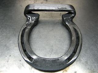
Patent Bar Horseshoe


The Patent bar horseshoe, is used to keep a horse from bearing weight on an injured leg. The shoe features a bar between the heels, which is raised 1 to 1½ inches to decrease the angle between the pastern and the cannon bone. In doing so, the shoe relieves stress on the flexor tendons and suspensory ligament.
 (The above shoe was constructed by David Boles and Marcus Lybarger)


The tenosynovitis imaged below is that of a horse with a severe injury to its deep digital flexor tendon in the pastern area (see ultrasound pic below). The horse was a grade 4/5 lame at the walk and was not willing to place his heel on the ground. By setting a patent bar shoe on the affected leg, the horse can bear weight on the limb with minimal strain on the injured deep digital flexor tendon. Over time, the heel elevation of the patent bar shoe is lowered until the horse is walking normally on the limb. This process usually takes 2-6 months depending on the extent of the injury. In addition, daily walking is encouraged to minimize the development of adhesions within the injured tendon/ligament. During the rehabilitation period, we recommend daily ice therapy, topical Surpass cream, and a series of extracorporeal shockwave treatment over the injured tendon/ligament. 


Severe tendinitis of the deep digital flexor tendon in the area of the pastern. The image on the left is a longitudinal view and the image on the right is a cross-sectional view of the same lesion. The dark or black area in the center of the image corresponds to fluid accumulation and fiber disruption (core lesion) within the body of the deep digital flexor tendon.