PHD Veterinary Service

PHD Veterinary Service

Contact Info

Dr. Porter @ 352-258-3571
portermi.dvm@gmail.com

Read more about Dr. Porter
And PHD Veterinary Services @


Showing posts with label lame horse. Show all posts

Friday, May 23, 2014

Severe Cartilage Degeneration in a Horse

A 12 year-old show jumper mare presented to PHD veterinary services for the history of recurring forelimb lameness. In the past 2 years, the mare had the left front coffin joint treated with corticosteroids and hyaluronic acid every 6 months. However, the most recent treatment with intra-articular corticosteroids did not result in resolution of the lameness. On presentation, the mare was a grade 3/5 lame in the left front. The lameness was most evident when trotting on firm ground and in a circle to the right. The mare was NOT positive to hoof tester exam and was mildly positive to lower limb flexion. The coffin joint was "blocked" with carbocain and the mare's lameness improved by >85%. Radiographic exam of the left front foot only revealed evidence of mild osteoarthritis of the coffin joint.  (Figure 1).
Figure 1
Due to the following factors: 1) a history of poor response to intra-articular therapy, 2) a positive response to blocking the coffin joint, and 3) minimal data via the radiographic exam, it was recommended that the distal forelimb of this mare be evaluated via magnetic resonance imaging (MRI).  The MRI exam requires general anesthesia and generally costs between $1800-$2000; however the information gathered from an MRI exam is far superior than any other type of diagnostic exam! As such, I strongly recommend considering an MRI exam in cases that the CAUSE for the lameness has not been determined and a reasonable attempt has been made to treat and/or diagnose the problem. Figures 2-4 includes several images from the MRI exam involving this show jumper mare. A small, blue circle is included in each image to high light the large articular cartilage and subchondral bone defect that was discovered through the MRI study. In each image, the lesion appears as a small black circle at the surface of the bone. In addition to this finding, the MRI study noted additional evidence of chronic degeneration of the coffin joint. The full extent of the degeneration and the cartilage damage was NOT evident in the radiographic exam!!

Figure 2

Figure 3

Figure 4
Although the prognosis was poor for return to soundness, the mare was treated aggressively with various regenerative therapies and extended rest. Unfortunately, the mare remained significantly lame and it was determined that humane euthanasia was the best option due to the relentless foot pain. A post-mortom exam was performed and a very large area of abnormal cartilage loss (blue circle in Figure 5) was noted that corresponded to the MRI findings. This case represents yet another exam of the importance for getting an answer as to WHAT is causing the problem such that a prognosis can be provided followed by a proper course of action.

Figure 5
In the state of Florida, the most sensitive MRI for imaging in horses is ONLY available at the University of Florida's College of Veterinary Medicine. In addition, the board certified radiologists at the Veterinary College of Veterinary Medicine are especially well trained for the evaluation of MRI studies in horses! For more information about their services please go to:

http://largeanimal.vethospitals.ufl.edu/services/diagnostic-imaging/


Friday, February 7, 2014

Foundered Horse with Inadequate Shoeing!

A 10 year-old Arabian cross gelding presented for shifting foot lameness and pounding digital pulses. There was no history of laminitis/founder. The horse was not willing to walk on concrete and was a grade 3/5 lame in both front feet on soft ground. Physical exam noted a prominent fat pad over the tail head and the development of a cresty neck! A radiographic exam was performed on both front feet to determine the severity of the gelding's condition.

Figure 1
Figures 1-3 correspond to the lateral projection of the right front limb. There is a clear explanation for the shifting leg lameness and pounding digital pulses. In Figure 2, the yellow dotted lines correspond to the dorsal hoof wall and the dorsal border of the coffin bone. In a normal horse, these two lines are parallel. However, in the case of laminitis/founder with coffin bone rotation the yellow lines are NOT parallel. The small blue triangles labelled A and B correspond to the angles of the yellow lines with the horizon (red line). In this horse, angle A does NOT equal angle B. These findings confirm the diagnosis of laminitis with coffin bone rotation. The difference between angle A and B corresponds to the degrees of coffin bone rotation and in this case that number was 10-12 degrees. The left front limb was similar.


Figure 2
In addition, the gelding's cranial to caudal balance is extremely poor. The yellow line in Figure 3 corresponds to the "center of rotation" and in a normal horse should divide the foot evenly in half. As such, there should be equal amount of ground contact (red lines) in front of the yellow line (cranial) and behind the yellow line (caudal). In this horse, the amount of ground contact behind the yellow line is nearly zero compared to that in front of the yellow line. This is due in part to the extremely long "toe" and the shoe placement. The extremely long toe and cranial shoe placement adds significant leverage on the toe which will aggravate the laminitis and increase the horse's level of pain!. Initial management of this case involved removing the shoes and significantly reducing the length of the toe. In addition, a blood sample was tested for insulin resistance and the horse was confirmed to be suffering from insulin resistance secondary to metabolic syndrome. The gelding was treated with the appropriate diet, stall confinement, pain medication, and medical management for insulin resistance. Stay tuned for follow-up.....

Figure 3


Friday, September 27, 2013

Hind Limb Proximal Suspensory Ligament Desmitis in a Horse

A 15 year-old mare presented for a 3 week history of rear-limb lameness that was associated with a "drop" of the rear fetlock joint. On presentation there was moderate swelling of the lower limb, just below the hock joint and the mare was lame at the walk. In addition, there was a 90 degree drop of the fetlock/pastern axis as noted in Figure 1.

Figure 1

The primary mechanism involved in "suspending" the fetlock joint and maintaining the proper fetlock/pastern axis is the suspensory ligament (Figure 2). The suspensory ligament originates just below the hock (red arrow) and initially is one structure (body) that travels down the back of the lower limb (yellow arrow). Approximately half way down the canon bone the suspensory ligament splits into a medial (inside) and lateral (outside) branch. The suspensory branches attach to the sesamoid bones which are located just behind and below the fetlock joint. As such, the suspensory ligament helps "suspend" the fetlock joint and a  proper fetlock/pastern axis.

Figure 2
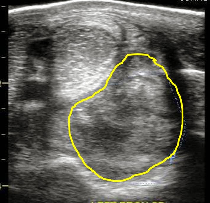
An ultrasound exam was performed to evaluate the entire suspensory ligament. The origin or proximal suspensory ligament is imaged in cross-section in Figures 3-6. The proximal suspensory ligament of the affected limb is grossly enlarged (yellow circle) and the fiber pattern is a mixed pattern with significant edema and evidence of active inflammation! There is a black and grey swirl pattern noted in the proximal suspensory ligament (tissue inside the yellow circle) of the affected limb which is indicative of severe changes.


Figure 3


Figure 4

 When compared to the normal limb, the significant increase in the size of the proximal suspensory ligament is evident. In this case the affected suspensory ligament was 2x the "normal" size. These ultrasound findings confirm the diagnosis of proximal suspensory desmitis of the hind limb. The prognosis for this injury is poor for return to riding and guarded for return to pasture soundness. Once the fetlock has "dropped" the physical changes to the suspensory ligament CANNOT be reversed!! Treatment is aimed at slowing the progress of the condition and attempting to provide pain relief to the horse. In my experience, corrective shoeing is the MOST important aspect of managing this condition.

Figure 5
  
Figure 6
A "fish tail" bar shoe is strongly recommended for this condition. The shoe should be set extra full such that approximately 1.5 to 2 inches of shoe extended BEHIND the heel bulb. Any kind of a wedge is CONTRAINDICATED in this condition! In addition, daily treatment with ice packs over the proximal suspensory ligament followed by topical Surpass cream are indicated to reduce inflammation and provide pain relief. With corrective shoeing, adequate pain relief, and supervised turn-out, these horses may return to pasture soundness however such a condition carries a guarded prognosis.

Figure 7


Friday, July 26, 2013

Plantar Annular Ligament Desmitis in a Horse


A 10 year old gelding presented for bilateral rear-limb lameness associated with excessive "wind puff" formation. The gelding typically had some wind puff formation above the rear fetlock joints but it had suddenly increased and subsequently the lameness was noted. Physical exam noted moderate to severe fluid accumulation within the distal flexor tendon sheaths, otherwise known as wind puffs (Figures 1-2). The fluid within the flexor tendon sheath tends to accumulate at the top of the sheath due to a very thin band of connective tissue that spans the back or plantar/palmar aspect of the fetlock. This tissue is called the plantar/palmar annular ligament (PAL) of the fetlock (Figure 2 (blue circle))

Figure 1
When fluid begins to accumulate within the tendon sheath due to simple tenosynovitis of the sheath or a tendon/ligament injury, the PAL becomes restrictive resulting in a "wind puff" (Figure 2, red circle), which consists of a pocket of fluid noted just above the fetlock joint. As the fluid increases in volume the PAL is stretched and inflammation develops within the ligament. Once inflammation develops within the PAL, lameness in that limb is common.

Figure 2
 Diagnosis is confirmed via ultrasonic exam. In Figures 3-4, the flexor tendons within the sheath are imaged and include the superficial flexor (SDF: blue) and deep digital flexor tendon (DDF: green). In addition, the PAL is visualized directly above the SDF tendon. Normally, the PAL is very thin and only measures 2-4mm in thickness. In this horse, the PAL measured nearly 10mm in thickness (blue arrows) and there were pockets of edema (black areas) along with poor fiber alignment! Chronic thickening of the PAL ligament may result in the inflammation within the SDF tendon which will significantly worsen the prognosis and lameness!  For this reason, careful examination via ultrasound is key.

 (Figure 3
 In this particular horse there does not appear to be involvement of the SDF or DDF tendons. The PAL appears very thick and inflamed. In Figure 4, the same area is imaged in a cross sectional view (left image) and a transverse view (right image). The significant amount of edema and disrupted fiber pattern within the PAL is quite evident in the both views!! Management of this condition involves corrective shoeing, topical anti-inflammatory application, and in my opinion surgical intervention. Once the PAL is grossly thickened, it is my experience that only surgical transection of the PAL ligament will provide a long term solution for the horse. It is very important to thoroughly ultrasound the limb through the pastern area to ensure that no ligaments or tendons are involved!! Finally, wind puffs are a common occurrence in many sport horses and usually indicate that the horse is in steady work. However, asymmetric wind puffs or a sudden increase in size is not normal and merits examination by your veterinarian!!

Figure 4

Friday, July 19, 2013

Thin Soles in a Horse!!


The radiographs below in Figures 1-3 belong to a middle-aged QH gelding that presented for severe lameness. The gelding was recently trimmed by the farrier and had a history of mild to moderate foot pain.
On presentation, the gelding was very sore when asked to walk on a firm surface and was not willing to walk in a tight circle! There were NO pounding digital pulses however firm pressure, via my finger tips, over the sole region resulted in a strong pain reaction by the gelding. Radiographs were taken and severely thin soles were diagnosed in both front feet (Figure 1 and 2). The actual thickness of the sole was difficult to measure due to the thinness but was approximated at 2-3mm (Figure 3). Less than 5mm of thickness essentially corresponds to 1 swipe with the hoof knife!!

Figure 1

Figure 2


Figure 3


In Figure 4, there is a radiograph of a normal horse's foot. The sole depth at the toe region is approximately 15-20mm. In my experience, when the sole thickness is less than 10mm, there is increased likelihood of foot pain from concussion with the ground. Thin soled horses are at high risk for sole bruising and sub-solar abscess development. Unfortunately, some horses are "naturally" thin soled due to poor genetics and MUST be managed with shoes. The corrective shoeing is simple and may include a rim pad, leather pad , or soft pour-in pad. I strongly recommend radiographic evaluation PRIOR to any foot trimming to determine the sole thickness of horses that have a history of chronic foot pain!

Figure 4

Friday, March 29, 2013

Coffin Bone De-Rotation!!

Approximately 6 weeks ago, a middle aged mare presented for chronic founder and non-weight bearing lameness. At presentation, there was significant rotation of the coffin bone in the lame foot (greater than 15 degrees) and reduction in the sole depth at the toe region (Figure 1-3). Due to the severe degree of coffin bone rotation and lameness it was determined that transection of the deep digital flexor tendon was the best option for the mare. The procedure was performed at the barn with sedation and a regional block.

Figure 1


Figure 2

Figure 3
In addition to transection of the deep digital flexor tendon, corrective shoeing was achieved by an expert farrier who specializes in foundered horses  (Sir Adam Whitehead). Corrective shoeing is as important if not MORE important than the surgical procedure and without it the procedure could not be a success! The mare's lameness has slowly improved over the past 4 weeks and follow up radiographs revealed complete DE-ROTATION of the coffin bone (Figure 4).  The palmar angle (blue line) is now approximately zero degrees with respect to the horizon and there has been a moderate increase in the sole depth at the toe region (yellow arrow).

Figure 4

In my experience, horses coffin bone rotation that exceeds 15 degrees benefit greatly from this procedure. Typically, these horses require 4-6 months from time of surgery before they are barefoot and sound in the pasture. This mare's initial recovery was slowed due to a sub-solar abscess that involved her entire sole and communicated with the coronary band. 

Thursday, February 21, 2013

Laminitis in a horse!!

 The following radiographs are from a gelding that has been lame for nearly 1 year. The gelding has been treated for laminitis/founder and despite efforts by the farrier and veterinarian, the gelding's conditions has worsened. He is non-weight bearing on one forelimb at the time of my exam. The right front foot is imaged in Figure 1 and the left front foot in Figure 2. In Figures 3 and 4 the palmar angle (angle "A") is measured in both front feet. The palmar angle corresponds to the angle that the bottom or palmar surface of the coffin bone makes with the horizon. In both front feet, the palmar angle measures approximate +23-25 degrees. Normally, the palmar angle may range between zero to +5 degrees. Abnormal palmar angles may be negative or greater than +7-8 degrees. However, even though the right and left front feet share the same palmar angle, it is for two very different reasons!

Figure 1 (Right front)

Figure 2 (Left front)
Figure 3 (Right front)
Figure 4 (Right front)
The most likely causes for an increased palmar angle include coffin bone rotation and coffin joint contracture. The difference between these two conditions is key to making the correct diagnosis. In the case of laminitis, the coffin bone rotates under the influence of the deep digital flexor tendon and due to a lack of attachment between the coffin bone and the lamina (i.e. laminitis). In Figure 5, the degree of coffin bone "rotation" is estimated based on the difference between angles "A" and "B". Normally, the hoof wall (red line over "A")  is parallel with the coffin bone (red line over "B"). When these two lines are parallel, the angles equal each other and there is zero degrees of coffin bone rotation. Therefore, the problem with the left front limb in Figure 5 is laminitis and secondary coffin bone rotation.

Figure 5 (Left front)
The right front foot has zero degrees of coffin bone rotation however the palmar angle is the same as in the left front foot. This due to coffin joint contracture and is the definition of a "Club foot". In Figure 6, the lines corresponding to the hoof wall and coffin bone are parallel and as such angle  "A" equals angle "B". Figure 7 diagrams the concept of coffin joint contracture that results in an angle of contracture (angle "A"). Typically, a club foot or coffin joint contracture does not result in non-weight bearing lameness but rather a life long commitment to proper shoeing and trimming. In this case, first glance of the radiographs might be deceiving because both front feet have an abnormal palmar angle, but it is the left front foot with the coffin bone rotation and life threatening laminitis! The deep digital flexor tendon was transected in the left front limb and follow-up radiographs will follow!
To be continued......

Figure 6 (Right front)
Figure 7 (Right front)


Thursday, February 7, 2013

Proximal Suspensory Desmitis in a Horse

The ultrasound images below are from a teenage gelding that presented for a 3 month history of mild, forelimb lameness. The lameness would improve with rest but would return soon after the gelding was returned to work. On physical exam, the proximal suspensory palpated sensitive, just below the carpus (knee) of the right forelimb. The gelding was not lame in a straight line however when lunged in a circle to the left, a mild lameness (2/5) was noted in the right forelimb. The lameness improved approximately 50% after blocking the lower limb however when the proximal suspensory ligament was blocked, the gelding was sound. Ultrasound exam of the tendons and ligaments revealed a focal area of decreased echogenicity (dark spot) which was consistent with inflammation and edema within the proximal suspensory ligament. 

Figure 1

Figure 2
In Figure 1, there is a cross sectional image of the superficial flexor tendon (SDF), deep digital flexor tendon (DDF), distal check ligament, and the proximal suspensory ligament. This image was made approximately 10cm below the knee or carpus. In Figure 2, the same area is imaged in cross section and in longitudinal plane. The same lesion (dark spot) can be seen in both images which is consistent with a "real" lesion versus an artifact. In the longitudinal view (right image in Figure 2), the origin of the suspensory is highlighted by the blue arrows and the edema is noted by the dark fibers just above the blue arrows. In Figure 3, the cross sectional image is slightly obliqued to visualize the inside or medial aspect of the limb. The lesion within the suspensory ligament is more apparent in this image and is represented by the dark blue circle within the yellow circle (suspensory ligament) in Figure 3.


Figure 3

Figure 4

The history and lameness exam findings are "classic" for a forelimb proximal suspensory ligament injury. Often the lameness is most noticeable when the affected limb is on the outside of the circle and the lameness will improve temporarily with rest. The prognosis for this injury is "good" however will require rest, ice therapy, corrective shoeing and a specific rehabilatory program. Adjunct therapies include shockwave treatment and platelet rich plasma (PRP) injections.


Thursday, December 27, 2012

Long toe/Under run heel!!

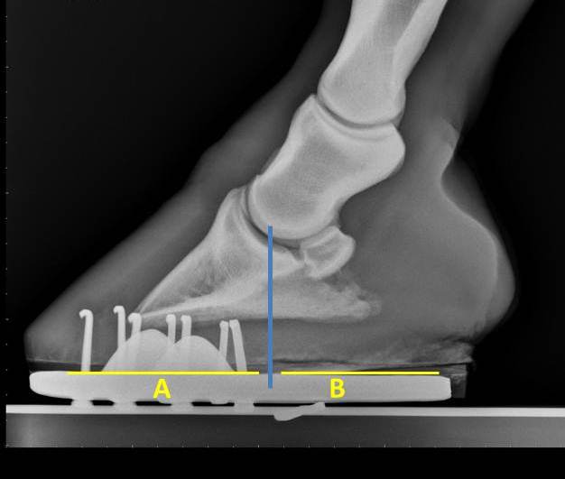
The image below is of a horse's foot that is suffering from a conformational problem known as "long toe-under run heels". This horse tends to grow significant amount of hoof wall within 4-5 weeks without growing normal heel. Although the heel regions grow, they tend to roll under the center of the hoof. As such, this horse and others like him are prone to heel pain and poor performance. In Figure 1 the center of rotation is marked by the blue line. Ideally, the length of foot that is in front of the blue line and behind should be equal. This would result in a horse that is balanced "cranial to caudal" with respect to the center of rotation. 


Figure 1

As the amount of foot behind  (B) or caudal to the center of rotation decreases compared to "A" there is  a significant increase in the amount of force that is applied across the heel regions. This results in the rolling under or "under run heels" that is imaged in Figure 1 and 2. When this horse is trimmed, the hoof wall needs to be trimmed back to the widest portion of the frog.  This may seem counter intuitive due to the apparent "lack of heel" however it is necessary to achieve "normal" heel growth. 
Figure 2

The radiograph below is that of horse that has heel pain and is lame when trotted in a small circle in both directions. The hoof tester exam noted a strong positive response across the bar region of both heels. Notice that the length of "B" is significantly shorter than "A". In order to correct this problem, corrective shoeing is a must!  The process begins with a balanced trim that includes lowering the heels to the widest portion of the frog. This is followed by applying a shoe that either has built-in break over such as a natural balance/equilibrium shoe or break over is increased manually be rolling the toe. In addition  the shoe is set extra full in an attempt to increase the length of "B" and hence support the caudal aspect of the foot. 
Figure 3

In Figure 4 the horse has been trimmed and re-set. Notice that the length of  "B" is nearly the same as "A". This horse is quite close to being balanced with regard to the center of rotation (blue line) and within 4-5 days returned to complete soundness. Finally, these horses that tend to have a long toes and under run heels need to be trimmed and re-set every 4-5 weeks. It is quite common that these horses are sound for the first 4 weeks after the farrier visit yet their performance begins to diminish as 5 to 6 weeks pass before they are trimmed and re-set. Although radiographs are not necessary to diagnose this problem, they are helpful for quantifying the extent of imbalance and documenting improvement after shoeing. 
Figure 4


Friday, May 11, 2012

Mesotherapy 


These small bumps were caused by micro-injections of medication just under the skin, or also known as Mesotherapy. This treatment has been used effectively for over 30 years in humans and horses. The primary objective of this treatment in horses is to block or break the pain cycle associated with chronic back pain. 

Technically, mesotherapy treatment stimulates the mesoderm or middle layer of skin by stimulating the giant fibers present within this layer of skin. Commonly, a combination of lidocain and dexamethasone is injected via multiple extra small needles (30g). These micro-injections result in stimulation of the giant nerve fibers located within this layer of skin that also communicate with major nerve tracts within the spinal chord. These nerve fibers INHIBIT the conduction of pain signals to the brain.

Typically, one or two mesotherapy treatments are applied to a horse's back and often are combined with shockwave treatments. Mesotherapy is used primarly as an aid to alleviate back pain followed by treatment of the primary cause of the back pain/spasm. This typically involves a source of lameness in either the forelimbs or hindlimbs. In addition, osteo-arthritis of the thoraco-lumbar spine and "kissing spines" are causes of chronic back pain. 

The response to mesotherapy is usually within 2-3 days and may last for 3-4 months.