PHD Veterinary Service

PHD Veterinary Service

Contact Info

Dr. Porter @ 352-258-3571
portermi.dvm@gmail.com

Read more about Dr. Porter
And PHD Veterinary Services @


Showing posts with label florida equine vet. Show all posts

Friday, September 26, 2014

Fungal Plaque in a Horse

A 7 year-old gelding presented to PHD veterinary services for the complaint of mild epistaxis (bloody nasal discharge). Endoscopic exam of the nasal passages identified a golden colored mass (Figure 1) that was covered in blood and was located within the opening to the ethmoid turbinates. The mass was diagnosed as an ethmoid hematoma based on location, appearance, and behavior. The client was given the option of surgical removal or treatment with intra-lesional doses of formalin. Based on the relatively small size of the ethmoid hematoma it was decided to attempt treating 1-2x with formalin and if there was not complete resolution then surgical resection would be pursued.


Figure 1
Four weeks after the first injection of formalin, a follow-up endoscopy noted significant reduction in the size of the ethmoid hematoma (Figure 2) and what appeared to be a second mass deeper within the ethmoid turbinates. The second ethmoid hematoma was not visualized during the initial exam because the first ethmoid hematoma was blocking the view! Based on these findings, the second ethmoid hematoma was injected with formalin in a similar fashion as the original ethmoid hematoma.

Figure 2
Six weeks after the second formalin injection the client reported that there was a slight yet persistent bloody discharge from the affected nares. Endoscopic exam revealed what appears to be a fungal plaque (black/white/yellow) adhered to the site of the ethmoid hematoma (Figure 3 and 4). This is an unusual finding and may prove to be a challenging complication. Fungal plaques have a predilection for vascular tissue and can infect the upper airway of horses. Fungal infection within the guttural pouch of a horse is well documented and can result in a catastrophic hemorrhage if not diagnosed and treated early. Fungal infection within the ethmoid turbinates is not common in my experience. Often these fungal plaques do NOT respond to systemic anti-fungal medication and must be either removed or treated aggressively with topical medication.
.
Figure 3

Figure 4
The current clinical plan for this horse is medication with oral anti-fungal medication for several weeks. If there is no change, the fungal plaques will be treated with topical anti-fungal medication. Stay tuned....

Finally after 4 weeks of anti-fungal medication, there is no evidence of a fungal plaque and there is NO evidence of an ethmoid hematoma (Figure 5). The ethmoid turbinates are visible for the first time in this horse since the initial exam. Although there is no evidence of an existing ethmoid hematoma, these tumors commonly reoccur hence the gelding will be monitored closely for the next 12 months. 


Figure 5

Saturday, August 16, 2014

Drug Testing and Prepurchase Exams for Horses

A 10 year old, warm-blood mare presented to PHD veterinary services for a prepurchase exam. The standard exam, complete with limb flexion was performed and the mare was found to be completely sound and negative to limb flexion. The buyer requested baseline radiographs of both hocks and both front feet. In addition, it was strongly recommended that a drug screen of the mare's blood be performed for the detection of sedatives, anti-inflammatory medications and corticosteroids.
.
.
.
Figure 1

Although the mare was sound and did not resent limb flexion, there was radiographic evidence of advanced osteoarthritis of the lower hock joints.  In Figures 1,2, and 4, the yellow arrows are pointing to radiographic changes consistent with arthritis along the front or dorsal aspect of the lower hock joints. There is new bone growth (osteophytes) and joint space narrowing. In addition, in Figure 3, the blue arrows are pointing to the area of sclerosis surrounding the lower hock joints which suggests chronicity of the arthritis and the apparent "fusion" of the distal hock joints. These radiographic changes are advanced and are surprising considering that the mare was sound for the prepurchase exam.

Figure 2

Figure 3
Even though the mare was sound, I did NOT pass this horse for sale and intended use based on radiographic findings and advised the buyer to at least wait for the drug screen results prior to making their decision!   The standard drug screen takes approximately 5-7 days for results to be reported. Interestingly, the mare's blood tested positive for high doses of an anti-inflammatory medication which likely explains why this horse was sound and negative to limb flexion even though she has advanced arthritis in the lower hock joints. This case represents yet another example of the importance of drug testing and base line radiographs for prepurchase exams!! Admittedly, it is a financial slippery slope once you begin the radiographic exam regarding how many areas to evaluate, however; areas of high probablity such as the hocks and front feet should always be considered!

Figure 4

Friday, April 25, 2014

Sarcoid Tumor in a Horse

A middle-aged gelding presented to PHD Veterinary services for the removal of a large tumor from the base of the right ear (Figures 1 and 2). Two years prior, a smaller tumor was surgically removed from the same ear. The histological report was consistent with a sarcoid tumor. At the time of the original surgery, several sarcoids were removed from different areas including the pectoral region and groin area. The recurring sarcoids were treated with intra-lesional chemotherapy, cryotherapy, topical medications and systemic herbal remedies. At presentation, a large, lobulated tumor was "hanging" from the base of the right ear. There was mild hemorrhage and evidence of moderate necrosis of the tumor. The gelding was resistant to manipulation of the tumor however gentle palpation of the ear noted that the tumor was NOT attached to the cartilage portion of the ear but only the skin and immediate subcutaneous tissues.

Figure 1


Figure 2

The gelding was rendered unconscious via injectable anesthetics and the tumor was removed through a large incision (Figure 3 and 4). Moderate hemorrhage was encountered confirming that the tumor was highly vascular. The incision was partially closed with sutures and the horse was recovered from anesthesia. Examination of the tumor after removal noted it consisted of 3 individual masses and weighed more than one pound (Figure 5).

Figure 3

Figure 4

Figure 5
This case represents an extreme example of recurrent sarcoids in a horse. Per the owner, the previous management of the sarcoids were initially effective however in time the tumors returned. The removal or de-bulking of the ear-based tumor will most likely not be sufficient in keeping the sarcoid tumor from removal. Aggressive therapy involving intra-lesional chemotherapy agents and cryotherapy will be employed to keep these tumors from continuing the develop!!


Figure 6
 Fast forward 6 weeks and the surgical wound is in the process of healing nicely (Figure 6), however there are several small sarcoids along the tip of the ear and also within the surgical site that are inflamed and beginning to grow. Follow-up treatment included the injection of cisplatin directly into the small sarcoids. Unlike the initial procedure which was done under anesthesia, the cisplatin injection was performed with sedation and a local "block" of the ear. Two weeks after the cisplatin injection, the small sarcoids along the tip of the ear are shrinking however the sarcoids within the surgical site remain inflamed  (Figure 7).

Figure 7
 Hence, the small sarcoids were treated with cryotherapy and the picture in Figure 8 was approximately 2 weeks post cryotherapy. The initial surgical site is a scar and all of the small sarcoids are regressing nicely. This horse will be monitored very carefully to make sure that any new sarcoid growth is treated aggressively!! This case represents an excellent example of the need for early, aggressive and multi-drug treatment of sarcoids in horses.
Figure 8

Friday, March 21, 2014

IIeal Hypertrophy in a Horse


A 10 year old Paso fino mare presented for recurrent colic for several months duration. The mare had a long history of being a "hard keeper" but in the past 3-4 months had begun to colic after each feeding. The mare was fed a diet of senior feed plus free-choice coastal bermuda grass hay. The mare was referred to PHD Veterinary services for a gastroscopy (stomach scope). The mare presented with a body condition score of 3 out of 9. The client reported that the mare had a good appetite yet shortly after eating, the mare would develop signs consistent with abdominal discomfort! The gastroscopy was normal therefore we opted to ultrasound the mare's abdomen.  In Figure 1 there is a cross-sectional image of a loop of intestine (black circle) that is grossly abnormal. The lumen (center) is completely filled in with soft tissue(grey area). The filled in center in Figure 1 corresponds to thickening of the small intestine which likely results in delayed passage of ingesta. Interestingly, there was only 1 very distinct area of the small intestine that scanned abnormally thick. However, there were several loops of small intestine that were dilated and had diminished peristalsis (Large black circles in Figure 2).


Figure 1
Figure 2

The ultrasound findings were consistent with a focal area of thickened small intestine and a large area of dilated small intestine. The dilated small intestine were likely "up stream" from the thickened small intestine and were as a result of partial obstruction of the thickened small intestine. Based on these findings, the mare was referred for abdominal exploratory surgery. During the abdominal exploration, a very thick region of the small intestine was identified. This region corresponded to the ileum which is the very final section of the small intestine. Approximately 12 inches of ileum were grossly thickened resulting in minimal lumen formation for passage of ingesta (Figure 3). Normally, the ileum is a wide-open tube as depicted in Figure 4!! Unfortunately, due to the severity of the condition, poor prognosis, and financial limitations, the mare was euthanized on the surgical table. Ileal hypertrophy has been reported in horses consuming coastal bermuda grass hay and it is theorized that some horses develop an "allergic" response to coastal hay resulting inflammation of the ileum and ultimately gross thickening. More commonly, colic symptoms associated with coastal hay is due to poor quality hay rather than ileal hypertrophy. However, any horse with chronic colic symptoms that is consuming coastal bermuda grass hay should be evaluated for this condition. 

Figure3


Figure4

Friday, March 7, 2014

Fractured Coffin Bone in a Horse

The radiographic images below are from a horse that presented for a 2 week history of non-weight bearing lameness in a forelimb. The horse had been treated for a suspect abscess with minimal clinical improvement. Interestingly, the horse was minimally responsive to hoof tester exam.

Figure 1
In the lateral radiograph (Figure 1) there is no obvious explanation for the lameness. However in the subsequent radiographs (Figures 2-4) the cause for the lameness is evident. A large fracture of the coffin bone is apparent. The fracture extends from the toe region all the way through the joint!

Figure 2
 In Figures 3 and 4, the fracture line appears to encompass the entire length of the coffin bone. The prognosis for future soundness depends on whether the fracture communicates with the coffin joint. If it does, the prognosis for soundness is poor due to the likelihood of developing of advanced osteoarthritis. This type of fracture will eventually heal with the correct shoeing and adequate rest. However, the prognosis for return to soundness is guarded due to the severity of this fracture!

Figure 3

Figure 4

Friday, February 7, 2014

Foundered Horse with Inadequate Shoeing!

A 10 year-old Arabian cross gelding presented for shifting foot lameness and pounding digital pulses. There was no history of laminitis/founder. The horse was not willing to walk on concrete and was a grade 3/5 lame in both front feet on soft ground. Physical exam noted a prominent fat pad over the tail head and the development of a cresty neck! A radiographic exam was performed on both front feet to determine the severity of the gelding's condition.

Figure 1
Figures 1-3 correspond to the lateral projection of the right front limb. There is a clear explanation for the shifting leg lameness and pounding digital pulses. In Figure 2, the yellow dotted lines correspond to the dorsal hoof wall and the dorsal border of the coffin bone. In a normal horse, these two lines are parallel. However, in the case of laminitis/founder with coffin bone rotation the yellow lines are NOT parallel. The small blue triangles labelled A and B correspond to the angles of the yellow lines with the horizon (red line). In this horse, angle A does NOT equal angle B. These findings confirm the diagnosis of laminitis with coffin bone rotation. The difference between angle A and B corresponds to the degrees of coffin bone rotation and in this case that number was 10-12 degrees. The left front limb was similar.


Figure 2
In addition, the gelding's cranial to caudal balance is extremely poor. The yellow line in Figure 3 corresponds to the "center of rotation" and in a normal horse should divide the foot evenly in half. As such, there should be equal amount of ground contact (red lines) in front of the yellow line (cranial) and behind the yellow line (caudal). In this horse, the amount of ground contact behind the yellow line is nearly zero compared to that in front of the yellow line. This is due in part to the extremely long "toe" and the shoe placement. The extremely long toe and cranial shoe placement adds significant leverage on the toe which will aggravate the laminitis and increase the horse's level of pain!. Initial management of this case involved removing the shoes and significantly reducing the length of the toe. In addition, a blood sample was tested for insulin resistance and the horse was confirmed to be suffering from insulin resistance secondary to metabolic syndrome. The gelding was treated with the appropriate diet, stall confinement, pain medication, and medical management for insulin resistance. Stay tuned for follow-up.....

Figure 3


Friday, January 3, 2014

Wood Splinter Foreing Body in a Horse

A young mare presented with a baseball-size swelling on the left side of her neck that was just below the jugular groove. Two weeks prior, the mare presented with a puncture wound in the same area and the referring veterinarian removed several splinters. The mare was treated with antibiotics and the wound healed slowly; however, purulent discharge developed recently and the mare was tender to manipulation of the swelling. An ultrasound exam was performed and several linear foreign bodies (yellow arrows) were noted (Figures 1-3).

Figure 1
The linear foreign bodies were approximately 1-2 cm deep in the tissue and ranged in length from 5mm to 20mm (yellow arrows). Most concerning was the close proximity of one linear foreign body with the carotid artery (red circle in Figure 1)!!
Figure 2

Figure 3


 A 6cm incision was made directly over the linear foreign bodies followed by careful dissection of the subcutaneous tissues (Figures 4 and 5). Although the ultrasound exam suggested that the foreign bodies were big and obvious, it was quite tedious to locate the wooden offenders. In this case, 2 weeks had passed since the mare had impaled herself and her body had begun the process of forming thick scar tissue around the splinters, like a cocoon. After what seemed hours, 5 wooden splinters (Figure 6) ranging in size were removed from the mare's neck!!


Figure 4


The wound was lavaged heavily and left open to heal by second intention. It is probable that some wood fragments remain within the wound however their size and the extensive surgical exposure should allow for adequate healing without complications.

Figure 5


Figure 6


Friday, November 1, 2013

Urinary Bladder Stone in a Horse

A 10 year-old gelding presented for a history of frequent urination and blood-tinged urine. The referring veterinarian had analyzed the urine and confirmed the presence of blood. In addition, a CBC and Chemistry was performed on blood collected from the gelding and all parameters were within normal limits. During my initial exam, I sedated the gelding and performed a rectal exam. A plumb-size structure was palpated within the urinary bladder and the gelding became increasingly agitated as the structure was manipulated. Trans-rectal ultrasound noted a 4cm, hyperechoic (bright white) structure within the lumen of the urinary bladder (Figure 1).

Figure 1
Subsequently, a cystoscopy was performed to identify the abnormal structure within the urinary bladder. Once visualized, the structure was identified as a urinary bladder calculi or stone (Figures 2-4). The stone was circular in dimension and the surface was very spiculated which was contributing to irritation of the urinary bladder wall and intermittent hemorrhage from the urinary bladder wall.

Figure 2

Figure 3

Figure 4
Following the cystoscopy, a trans-abdominal ultrasound was performed on the left and right kidneys. Unfortunately, a 5cm stone was visualized within the renal pelvis of the left kidney (Figure 5). Surrounding the hyperechoic (bright white) line is black fluid which most likely consists of urine and a dilated renal pelvis. It is quite likely that this kidney is not filtering blood as it should and nearly non-functional. Fortunately, the right kidney was normal and the horse will be okay as long as the right kidney does not develop a problem!!

Figure 5
The gelding was referred to a surgical facility and the urinary bladder stone was removed successfully. However, the gelding will need regular monitoring for changes in renal function and the presence of new bladder/kidney stones!! The development of kidney/bladder stones in horses is random however horses that consume a diet rich in calcium may be at higher risk.

Friday, September 27, 2013

Hind Limb Proximal Suspensory Ligament Desmitis in a Horse

A 15 year-old mare presented for a 3 week history of rear-limb lameness that was associated with a "drop" of the rear fetlock joint. On presentation there was moderate swelling of the lower limb, just below the hock joint and the mare was lame at the walk. In addition, there was a 90 degree drop of the fetlock/pastern axis as noted in Figure 1.

Figure 1

The primary mechanism involved in "suspending" the fetlock joint and maintaining the proper fetlock/pastern axis is the suspensory ligament (Figure 2). The suspensory ligament originates just below the hock (red arrow) and initially is one structure (body) that travels down the back of the lower limb (yellow arrow). Approximately half way down the canon bone the suspensory ligament splits into a medial (inside) and lateral (outside) branch. The suspensory branches attach to the sesamoid bones which are located just behind and below the fetlock joint. As such, the suspensory ligament helps "suspend" the fetlock joint and a  proper fetlock/pastern axis.

Figure 2
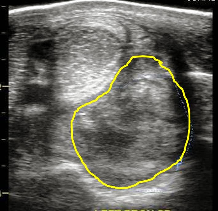
An ultrasound exam was performed to evaluate the entire suspensory ligament. The origin or proximal suspensory ligament is imaged in cross-section in Figures 3-6. The proximal suspensory ligament of the affected limb is grossly enlarged (yellow circle) and the fiber pattern is a mixed pattern with significant edema and evidence of active inflammation! There is a black and grey swirl pattern noted in the proximal suspensory ligament (tissue inside the yellow circle) of the affected limb which is indicative of severe changes.


Figure 3


Figure 4

 When compared to the normal limb, the significant increase in the size of the proximal suspensory ligament is evident. In this case the affected suspensory ligament was 2x the "normal" size. These ultrasound findings confirm the diagnosis of proximal suspensory desmitis of the hind limb. The prognosis for this injury is poor for return to riding and guarded for return to pasture soundness. Once the fetlock has "dropped" the physical changes to the suspensory ligament CANNOT be reversed!! Treatment is aimed at slowing the progress of the condition and attempting to provide pain relief to the horse. In my experience, corrective shoeing is the MOST important aspect of managing this condition.

Figure 5
  
Figure 6
A "fish tail" bar shoe is strongly recommended for this condition. The shoe should be set extra full such that approximately 1.5 to 2 inches of shoe extended BEHIND the heel bulb. Any kind of a wedge is CONTRAINDICATED in this condition! In addition, daily treatment with ice packs over the proximal suspensory ligament followed by topical Surpass cream are indicated to reduce inflammation and provide pain relief. With corrective shoeing, adequate pain relief, and supervised turn-out, these horses may return to pasture soundness however such a condition carries a guarded prognosis.

Figure 7


Friday, September 13, 2013

Fractured Patella in a Horse

A teenage gelding presented for sudden non-weight bearing lameness in the right hind limb. There was a history of a "kick" from another horse but the exact location of the kick was not witnessed. On presentation, there was a basketball-size swelling centered on the stifle and the horse was very painful. He was not willing to walk on the limb. The initial radiographs were not conclusive due to the severe swelling and a follow-up exam was performed 10 days later. On presentation, the gelding was walking on the limb but was very resistant to have the limb flexed at the stifle/hock. There was minimal swelling compared to previous exam.
Figure 1 is a lateral radiograph of the patella. The yellow lines corresponds to bone fragments that are noted along the edges of the patella.


Figure 1
In order to better assess the patella, a special "sky line" projection is required and was especially difficult in this horse because he was resistant to having the stifle flexed! However, it proved to be the most important radiographic view. In  Figure 2, the patella is imaged and a distinct line can be noted traveling through the body of the patella. In Figure 3, the "line" is highlighted in red and is consistent with a complete or near-complete fracture of the patella. In Figure 4, a normal patella is imaged for comparison to the fractured patella.

Figure 2


Figure 3


The degree of fracture appears complete or near-complete which significantly worsens the prognosis. There are cases of patella fracture in horses that are described in the literature that have healed after several months of stall rest. However, there is minimal discussion regarding the future of these horses with regards to as equine athletes. The prognosis for this horse to return to work is poor and his outcome is yet to be determined.