PHD Veterinary Service

PHD Veterinary Service

Contact Info

Dr. Porter @ 352-258-3571
portermi.dvm@gmail.com

Read more about Dr. Porter
And PHD Veterinary Services @


Showing posts with label horse ultrasound. Show all posts

Friday, January 31, 2014

Hip Subluxation in a Horse


A 10 year-old mare presented for sudden onset hind limb lameness. The mare was non-weight bearing on the right hind limb and within 24 hrs there was significant swelling centered over the right gluteal region. In Figure 1, the pelvis of a horse is pictured to identify the left (bottom blue arrow) and the right (top blue arrow) coxofemoral joints. In Figure 2 and 3, the pelvis has been rotated slightly to picture the area that is most commonly ultrasounded to assess the coxofemoral joint space. In Figure 3, the red line corresponds to the junction of the pelvis and the femor. The blue arrow corresponds to the coxofemoral joint space.

Figure 1

Figure 2


Figure 3
Figures 4 and 5 illustrate the views captured via ultrasound exam of the equine pelvis. This area is very difficult to adequately radiograph in a standing horse hence the ultrasound exam provides an opportunity to assess the coxofemoral joint space of a horse. As in Figure 3, the red line in Figure 5 corresponds to the juction of the pelvis and femur (coxofemoral joint space) which is also highlighted by the blue arrow. In the normal exam, the only bone structures commonly noted are the pelvis (long white line at bottom of view) and the greater trochanter of the femur (small white line at the top right of the image).
Figure 4

Figure 5
Figures 6 and 7 correspond to the right coxofemoral joint space of the lame horse. There is clearly and extra curved line in the image that corresponds to the "head" of the femur. Normally, the head of the femur is NOT visualized because it is within the acetabulum that makes up the coxofemoral joint. The ultrasound findings below are consistent with a subluxation of the coxofemoral joint space and very likely fracture of the acetabulum!  This injury carries a very poor prognosis for an adult horse; however, in miniature horses or very small ponies, surgical correction exists as an option with a guarded prognosis.

Figure 6


Figure 7


Friday, September 27, 2013

Hind Limb Proximal Suspensory Ligament Desmitis in a Horse

A 15 year-old mare presented for a 3 week history of rear-limb lameness that was associated with a "drop" of the rear fetlock joint. On presentation there was moderate swelling of the lower limb, just below the hock joint and the mare was lame at the walk. In addition, there was a 90 degree drop of the fetlock/pastern axis as noted in Figure 1.

Figure 1

The primary mechanism involved in "suspending" the fetlock joint and maintaining the proper fetlock/pastern axis is the suspensory ligament (Figure 2). The suspensory ligament originates just below the hock (red arrow) and initially is one structure (body) that travels down the back of the lower limb (yellow arrow). Approximately half way down the canon bone the suspensory ligament splits into a medial (inside) and lateral (outside) branch. The suspensory branches attach to the sesamoid bones which are located just behind and below the fetlock joint. As such, the suspensory ligament helps "suspend" the fetlock joint and a  proper fetlock/pastern axis.

Figure 2
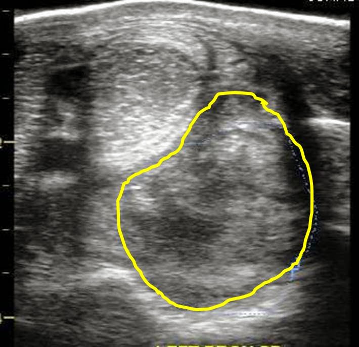
An ultrasound exam was performed to evaluate the entire suspensory ligament. The origin or proximal suspensory ligament is imaged in cross-section in Figures 3-6. The proximal suspensory ligament of the affected limb is grossly enlarged (yellow circle) and the fiber pattern is a mixed pattern with significant edema and evidence of active inflammation! There is a black and grey swirl pattern noted in the proximal suspensory ligament (tissue inside the yellow circle) of the affected limb which is indicative of severe changes.


Figure 3


Figure 4

 When compared to the normal limb, the significant increase in the size of the proximal suspensory ligament is evident. In this case the affected suspensory ligament was 2x the "normal" size. These ultrasound findings confirm the diagnosis of proximal suspensory desmitis of the hind limb. The prognosis for this injury is poor for return to riding and guarded for return to pasture soundness. Once the fetlock has "dropped" the physical changes to the suspensory ligament CANNOT be reversed!! Treatment is aimed at slowing the progress of the condition and attempting to provide pain relief to the horse. In my experience, corrective shoeing is the MOST important aspect of managing this condition.

Figure 5
  
Figure 6
A "fish tail" bar shoe is strongly recommended for this condition. The shoe should be set extra full such that approximately 1.5 to 2 inches of shoe extended BEHIND the heel bulb. Any kind of a wedge is CONTRAINDICATED in this condition! In addition, daily treatment with ice packs over the proximal suspensory ligament followed by topical Surpass cream are indicated to reduce inflammation and provide pain relief. With corrective shoeing, adequate pain relief, and supervised turn-out, these horses may return to pasture soundness however such a condition carries a guarded prognosis.

Figure 7


Friday, June 14, 2013

Shoulder swelling in a Horse

The mare in Figure 1 presented for a complaint of sudden onset severe swelling at the point of the right shoulder and near non-weight bearing lameness of the same limb. The mare is normally kept with other horses and there is no history of a "equine dispute" or evidence of external trauma. On presentation, the swelling consists of significant edema and the mare is considerably "tender" to pressure and palpation of the swelling. Manipulation of the limb was limited due to the significant pain response that resulted. Radiographic exam of the horse's shoulder is a viable option however due to the pain on manipulation and significant soft tissue swelling, an ultrasound exam was more highly indicated.

Figure 1
The images in Figure 2 and 3 correspond to the soft tissue swelling noted in Figure 1. A large, fluid filled pocket was noted deep to the skin surface. The fluid had a mixed pattern of echogenicity suggesting that the fluid consisted of cellular debris mixed with blood/serum. These findings are most consistent with a large subcutaneous abscess. The two most likely causes for abscess formation in this area would be trauma induced or abscess formation from a bacteria called corynebacterium pseudotuberculosis. This condition is also known as "Pigeon's fever". Pigeon's fever is a serious condition due to the highly infectious nature of the bacteria AND the fact that once the bacteria is present on the premises, it is very difficult to remove from the environment. Correct diagnosis depends on collecting a sample of fluid from the abscess and submitting for laboratory evaluation. Treatment of either condition requires establishing drainage of the abscess and daily  lavage for 10-14 days after drainage. Microbiological identification in this case is pending and the horse is in quarantine on the farm! The abscess was opened and drainage established.

Figure 2


Figure 3
Interestingly, within 7 days of examining the mare above, a gelding presented for similar symptoms. He was found non-weight bearing lame in the pasture with severe swelling of the shoulder region. The referring veterinarian provided digital radiographs which did not identify the cause of the lameness/swelling. On ultrasound exam, a bright, bone-like structure (red dotted line) was noted extending into a fluid filled mass that appeared to be a hematoma (Figure 4 and 5). The bone-like structure was the humerus bone (red dotted line) and it was fractured plus displaced. This gelding was humanely  immediately euthanized!

Figure 4

Figure 5
Although these cases are quite different in outcome, they demonstrate the benefit of soft tissue ultrasound with regards to assessing "swellings". As demonstrated it is possible to evaluate soft tissue changes as well as bony changes.


Thursday, May 3, 2012

Asymmetric pelvis


The pictures above are those of a horse with an asymmetric pelvis. Specifically, the left tuber sacrale is lower than the right. Less severe varieties of this problem have been described as "hunters bumps". The appearance of asymmetric tuber sacrale or "hunters bump" is in indication of a pathological problem within the pelvis/sacrum area. In the case above, the gelding suffered a fractured pelvis that was diagnosed via ultrasound. This patient presented for non-weight bearing lameness however many with only mild changes do not present for lameness but rather "poor" performance.


In the image above, the left gluteal region was examined via ultrasound and an irregular margin was noted along the body of the ileum. Note the "step" along the bony margin in the pic below.


Less severe cases involve inflammation within the sacro-iliac joint and/or inflammation of the dorsal sacroiliac ligaments. These areas should be assessed for evidence of inflammation and treatment may include shockwave treatment, mesotherapy, and/or sacro-iliac joint injections.