PHD Veterinary Service

PHD Veterinary Service

Contact Info

Dr. Porter @ 352-258-3571
portermi.dvm@gmail.com

Read more about Dr. Porter
And PHD Veterinary Services @


Friday, October 25, 2013

Severe Roaring in a Horse!!

A teen-age mare presented for respiratory noise during exercise and poor performance. The mare had made noise during exercise for the past 2-3 years WITHOUT poor performance. Interestingly, the mare has always been resistant when cantering to the RIGHT! Recently, it was noted that the mare demonstrated signs of exercise intolerance at the canter. During the exam, the mare was asked to canter on a lunge line and she carried her head to the outside of the circle with her nose turned up. After 1-2 minutes of cantering to the right, it became increasingly more difficult for her to catch her breath and she would occasionally cough/gag violently!. 

Figure 1
 The mare was scoped immediately after exercise and complete paralysis of the left arytenoid was discovered (Figures 1-2). During inspiration, the left arytenoid was across mid-line (yellow line) and obstructing at least 50% of the mare's airway (Figure 2).  It is highly likely that during heavy exercise, the increased negative pressure during inspiration would cause the paralyzed arytenoid to completely cover the airway and prevent the mare from breathing! 

Figure 2
In addition, while scoping the horse, the mare tended to displace her soft palate in such a position that the epiglottis was trapped below (Figures 3-4). When this occurred, the mare instantly began to cough/gag until she corrected the displacement! Displacement of the soft palate is not always associated with arytenoid hemipligia but when it does occur may worsen the exercise intolerance.


Figure 3


The prognosis for this mare is dependent on additional evaluation by an equine surgeon and the recommended surgical procedure. If she is a good candidate for surgery and the surgery is a success, there is a good likelihood that she will return to work and perform significantly better than before.


Friday, October 18, 2013

Melanoma in a Horse

The images below are from a 20 year-old gelding that presented for the complaint of straining to defecate. Examination of the horse's perineum revealed multiple, lobulated soft tissue masses that were surrounding the anus and under-side of the tail head region (Figures 1 and 2). These soft tissue masses are consistent with cutaneous melanomas in a horse.

Figure 1

Figure 2
In addition, the owner had reported that the horse appeared to be having difficulty seeing from the right eye. Careful exam of both eyes noted the presence of a soft tissue mass within the anterior chamber of the right eye (Figure 3). Although this mass was not biopsied for obvious reasons, it is likely that the mass is a melanoma. Scientific studies report that approximately 14% of cutaneous tumors in horses are melanomas making them relatively common, especially in grey horses. In addition, studies report that 80% of grey horses that are over the age of 15 have multiple melanomas.

Figure 3
The most common site for melanomas is under the tail head and perianal region. Less common sites include base of the ear, periorbital (around or near the eye), lips, and lower limbs. Diagnosis is typically made by simple physical exam however some melanomas grow below the skin surface and a biopsy may be necessary to confirm the diagnosis. Unfotunately, melanomas continue to grow in size and multiply in number. As that happens, melanomas may become an significant problem as in this case in which they are obstructing the horse's anus. If diagnosed early, complete surgical resection is strongly recommended! Alternative therapies include intra-lesional chemotherapy treatment and systemic treatment with cimetadine.

Friday, October 11, 2013

Respiratory Disease in a Horse

A 12 year-old mare presented for a history of increased respiratory rate and effort during the late summer months. In addition, a white discharge was noted from both nares. At presentation the mare had labored breathing, significantly nasal flaring and there was a large abdominal component to her respiratory effort (see videos below). Auscultation of the lungs noted wheezes and crackles and a very mild cough was elicited when the larynx was palpated. The client was of limited financial means and asked that I perform the fewest tests necessary to determine the source of the problem! As such, we opted to begin with an upper airway endoscopy to determine if there was any abnormality within the larynx and if not take a look down the upper trachea. The larynx was normal however there was a significant amount of white debris within the upper trachea (Figures 1-2). This white debris is otherwise known as sputum and is produced within the lungs by inflammatory cells!


Figure 1

Figure 2
The sputum present within the trachea may be due to an allergic airway condition known as heaves/COPD or it may be due to broncho-pneumonia!  These conditions present with similar symptoms but are treated quite differently. Hence making the correct diagnosis is essential! There are several diagnostic tests which will evaluate the lungs and the fluid within the lungs however the one exam that will give you all the information necessary to determine the cause of the sputum and how to treat is a trans-tracheal wash.  The trans-tracheal wash is performed by catheterizing the trachea through a small incision that is made along the ventral aspect of the neck, approximately half way between the horse's throat latch and thoracic inlet. The catheter is passed into the trachea and down to the level of the lungs. A small volume of sterile saline is flushed into the lungs and then aspirated out for analysis. This sample of fluid should contain the sputum noted in the endoscopy exam. The sputum is analyzed for cell type and for the presence of bacteria. If bacteria are found then the sample is tested for which antibiotic is most ideal to treat the pneumonia. This procedure is easily performed in the field with mild sedation and proper technique!


In this case, the mare was noted to be suffering from heaves AND pneumonia. This is not common but does occur and requires very specific medication to save the horse's life!! The horse was treated with a combination of systemic corticosteroids and appropriate antibiotics. Clinically, the horse presented as a typical heaves case however if we had only treated with corticosteroids the broncho-pneumonia would have worsened significantly and potentially caused the death of the patient!!

Thursday, October 3, 2013

PHD Veterinary Service App


Take a picture of this QR code with your smart phone's QR code scanner (free app that is easily down loaded from your favorite app store). Once you scan the QR code select the option to open the App in Safari and then save to your phone's/pad's home screen.


The PHD Veterinary Service App will make it easier for you to track Dr. Porter as he travels throughout the State of Florida and southeastern Georgia. In addition, there are quick links to PHD Veterinary Service's Facebook page and Blog

Friday, September 27, 2013

Hind Limb Proximal Suspensory Ligament Desmitis in a Horse

A 15 year-old mare presented for a 3 week history of rear-limb lameness that was associated with a "drop" of the rear fetlock joint. On presentation there was moderate swelling of the lower limb, just below the hock joint and the mare was lame at the walk. In addition, there was a 90 degree drop of the fetlock/pastern axis as noted in Figure 1.

Figure 1

The primary mechanism involved in "suspending" the fetlock joint and maintaining the proper fetlock/pastern axis is the suspensory ligament (Figure 2). The suspensory ligament originates just below the hock (red arrow) and initially is one structure (body) that travels down the back of the lower limb (yellow arrow). Approximately half way down the canon bone the suspensory ligament splits into a medial (inside) and lateral (outside) branch. The suspensory branches attach to the sesamoid bones which are located just behind and below the fetlock joint. As such, the suspensory ligament helps "suspend" the fetlock joint and a  proper fetlock/pastern axis.

Figure 2
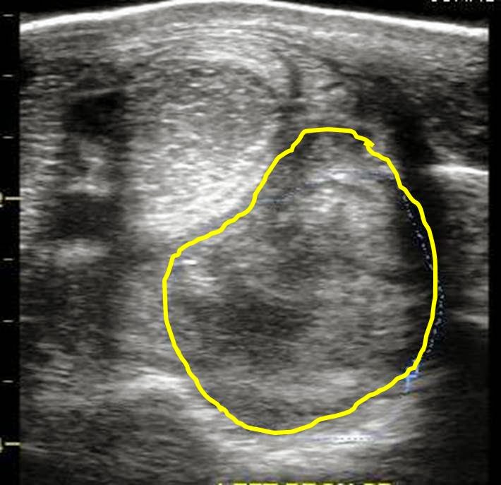
An ultrasound exam was performed to evaluate the entire suspensory ligament. The origin or proximal suspensory ligament is imaged in cross-section in Figures 3-6. The proximal suspensory ligament of the affected limb is grossly enlarged (yellow circle) and the fiber pattern is a mixed pattern with significant edema and evidence of active inflammation! There is a black and grey swirl pattern noted in the proximal suspensory ligament (tissue inside the yellow circle) of the affected limb which is indicative of severe changes.


Figure 3


Figure 4

 When compared to the normal limb, the significant increase in the size of the proximal suspensory ligament is evident. In this case the affected suspensory ligament was 2x the "normal" size. These ultrasound findings confirm the diagnosis of proximal suspensory desmitis of the hind limb. The prognosis for this injury is poor for return to riding and guarded for return to pasture soundness. Once the fetlock has "dropped" the physical changes to the suspensory ligament CANNOT be reversed!! Treatment is aimed at slowing the progress of the condition and attempting to provide pain relief to the horse. In my experience, corrective shoeing is the MOST important aspect of managing this condition.

Figure 5
  
Figure 6
A "fish tail" bar shoe is strongly recommended for this condition. The shoe should be set extra full such that approximately 1.5 to 2 inches of shoe extended BEHIND the heel bulb. Any kind of a wedge is CONTRAINDICATED in this condition! In addition, daily treatment with ice packs over the proximal suspensory ligament followed by topical Surpass cream are indicated to reduce inflammation and provide pain relief. With corrective shoeing, adequate pain relief, and supervised turn-out, these horses may return to pasture soundness however such a condition carries a guarded prognosis.

Figure 7


Thursday, September 19, 2013

Pyloris of a Horse of course!!

The endoscopic images in the following figures are of a horse's pyloris. This is the portion of the stomach that creates a valve and allows gastric fluids and contents to enter the small intestines. The very first region of the small intestines is known as the duodenum. In Figure 1, the pyloric sphincter is open and a small amount of feed material is noted around the edges of the sphincter. In Figure 2, the pyloris is nearly completely closed. The movement of material through the pyloris is controlled via rhythmic contractions or peristalsis of the stomach's muscular wall.

Figure 1
Figure 2
In Figure 3-6, there are very important abnormalities noted around the pyloric sphincter. In Figure 3 and 4, there is evidence of active bleeding from ulcerations around the pyloric sphincter! These ulcerations are likely to be a source of significance discomfort in the horse and would contribute to signs of gastric ulcer disease. It is possible and common to examine a horse's stomach and miss these lesions if the horse is not properly fasted prior to the gastroscopy and the pyloric sphincter is not visualized. This can be quite difficult in some equine patients!! The pyloric sphincter essentially controls the rate of gastric emptying hence any inflammation in this area will likely SLOW the rate of gastric emptying and result in an abnormal build-up of gastric fluid/content within the stomach.

Figure 3

Figure 4
 In Figures 5-6, there is a significant amount of hyperemia around the pyloric sphincter which corresponds to active inflammation. The degree of clinical signs may vary significantly from horse to horse however it is highly likely that horses with these types of lesions will have clinical signs consistent with gastric ulceration and WILL require the proper medical management! These type of lesions may be caused by excessive use of non-steroidal anti-inflammatory agents such as banamine and phenylbutazone. Diagnosis is dependent on a COMPLETE gastroscopy and I typically recommend a follow-up gastroscopy after treatment to verify complete resolution of the pyloric sphincter ulcerations!!

Figure 5

Figure 6


Friday, September 13, 2013

Fractured Patella in a Horse

A teenage gelding presented for sudden non-weight bearing lameness in the right hind limb. There was a history of a "kick" from another horse but the exact location of the kick was not witnessed. On presentation, there was a basketball-size swelling centered on the stifle and the horse was very painful. He was not willing to walk on the limb. The initial radiographs were not conclusive due to the severe swelling and a follow-up exam was performed 10 days later. On presentation, the gelding was walking on the limb but was very resistant to have the limb flexed at the stifle/hock. There was minimal swelling compared to previous exam.
Figure 1 is a lateral radiograph of the patella. The yellow lines corresponds to bone fragments that are noted along the edges of the patella.


Figure 1
In order to better assess the patella, a special "sky line" projection is required and was especially difficult in this horse because he was resistant to having the stifle flexed! However, it proved to be the most important radiographic view. In  Figure 2, the patella is imaged and a distinct line can be noted traveling through the body of the patella. In Figure 3, the "line" is highlighted in red and is consistent with a complete or near-complete fracture of the patella. In Figure 4, a normal patella is imaged for comparison to the fractured patella.

Figure 2


Figure 3


The degree of fracture appears complete or near-complete which significantly worsens the prognosis. There are cases of patella fracture in horses that are described in the literature that have healed after several months of stall rest. However, there is minimal discussion regarding the future of these horses with regards to as equine athletes. The prognosis for this horse to return to work is poor and his outcome is yet to be determined.

Thursday, September 5, 2013

Resolved Fibrosarcoma in a horse!

Several months ago I presented a case involving a gelding that presented for lameness and soft tissue swelling along the outside of the right knee (carpus). The gelding was lame at the walk and hesitant to flex the limb at the carpus. Ultrasound exam revealed a well demarcated soft tissue mass (dark tissue inside blue circle) that was centered over the carpus (Figures 1-3). The histopathology report was consistent with a fibrosarcoma.

Figure 1
Because the tumor was located immediately adjacent to the joint capsule of the carpus, surgical removal was considered high risk. As such, the tumor was injected with a chemotherapy agent called Cisplatin via ultrasound guidance. The tumor was injected twice, approximately 30 days apart. There was a moderate reduction in the size of the tumor just 3 weeks after the first injection and the gelding was no longer lame at the walk and was more willing to flex the limb at the carpus.

Figure 2


Figure 3
 The gelding's limb was evaluated 4 weeks after the second Cisplatin treatment. There was no evidence of the fibrosarcoma on the outside of the carpus AND under the skin (Figure 5). The ultrasound exam only noted normal subcutaneous tissue and joint capsule between the ultrasound probe and the carpus. The gelding was sound at the walk, trot, canter and has returned to full work!! The case is a good example of the benefits of intra-lesional injection of tumors that are non-operable with chemotherapy agents. There are several chemotherapy agents available and the procedure can be performed at the barn with ultrasound guidance.

Figure 4

Friday, August 30, 2013

Pleuropneumonia in a Horse

A 17 year-old gelding QH presented for a 3 week history of intermittent fever and coughing.

History:

The gelding had been treated with 3 different antibiotics over the past 3 weeks and there were no improvements in clinical signs. The problem started after the horse had been transported approximately 12 hours via truck and trailer. Within 24 hours of arriving to the show grounds the gelding was sick!

Physical exam:

When I examined the horse, the gelding was in distress! His heart rate was 80 bpm, respiratory rate was 60-70 bpm (shallow) and his body temperature was 102 degrees. Auscultation of the thorax noted lung sounds (sound of air moving in and out of lungs) on both sides of the horse only ABOVE the level of the shoulder but lung sounds were absent or muffled below the level of the shoulder. Suspecting pleuropneumonia, I performed a trans-thoracic ultrasound exam. A significant amount of fluid was noted in the pleural space along with large fibrin tags and multiple abscesses! ( Figure 1-3).

     
Figure 1

 In the video clips below, the fibrin tags can be seen "floating" in the excessive fluid within the pleural space. In addition, the fluid appears as "cellular" suggesting a heavy component of fibrin and purulent debris (pus).


In Figure 2-3, large abscesses are noted adjacent to the body wall. Ultimately, these abscess would need to be exteriorized through a rib resection in order for the horse to completely heal. Unfortunately, the ultrasound findings combined with the severe physical distress were very poor prognostic indicators and the owner elected humane euthanasia!  There was near zero chance that this horse could have been saved regardless of the medical and surgical intervention provided!

Figure 2


Figure 3
Pleuropneumonia in also called pleurisy and refers to bacterial infection of the pleural cavity and the surrounding soft tissue structures. This condition is VERY deadly and originates from bacterial colonization of the lower airway! The condition is more common in young horses that are transported long distances on a regular basis. Hence the term "shipping fever". A common belief is that horses which are transported with their head tied and a bag of hay in front of them are predisposed because they are not able to properly clear their airway of airborne debris and pathogens. Normally, horses eat with their head down which minimizes the passage of unwanted matter down their trachea and into their lungs.  This condition can be treated effectively if diagnosed early, i.e, within 1-2 days of clinical signs! Aggressive medical treatment is a must and includes IV antibiotics, chest drainage/lavage, and supportive care. One of the most painful horses I witnessed as a resident was a young filly with pleurisy!! Looked like a bad case of colic but was in fact pleuropneumonia!!


Friday, August 23, 2013

Aortic Valve Insufficiency in a Horse

Figure 1

Figure 2A and 2B
 The image in Figure 1 is of a horse's heart taken via trans-thoracic ultrasound. Specifically, the ultrasound probe is centered on the aortic valve. The horse presented for a prepurchase exam and a loud murmur was detected during the physical exam. There was no history of exercise intolerance nor was there evidence of heart failure during the physical exam. The murmur was best described as diastolic (during the filling phase) and included a high musical pitch at the end of the murmur. During the cardiac ultrasound exam, color flow doppler was imaged (Figure 1) to determine the flow of blood across the aortic valve. Normally, the color of the blood flow will be blue or red depending on whether the blood is moving towards or away from the ultrasound probe. When there is a "leaky" valve or a valve that is "insufficient" the color of the blood flow is bright yellow, white or orange. This color flow pattern is consistent with turbulence across the valve due to blood traveling "backwards" through the valve. In Figure 2, the red arrow identifies the normal direction of blood flow through the aortic valve. In Figure 3 a small white line is highlighted by the yellow arrows. This white line corresponds to one of the 3 valves that make up the aortic valve. In this horse, the aortic valve is thickened resulting in a "leaky" valve. The "leakiness" can be noted in Figure 4 as bright yellow and white blood flow in the opposite direction of normal blood flow across the aortic valve. The thickened valve likely vibrates as the jet of blood flows "backwards" causing the musical heart murmur!  Aortic insufficiency is the most common type of heart murmurs in horses and is often benign during the early phases of development. However, if the horse lives long enough, the aortic insufficiency can lead to heart failure in the horse. As such, heart murmurs in horses should not be dismissed as "normal" and inconsequential but should be carefully documented via ultrasound exam!!


Figure 3


Figure 4