PHD Veterinary Service

PHD Veterinary Service

Contact Info

Dr. Porter @ 352-258-3571
portermi.dvm@gmail.com

Read more about Dr. Porter
And PHD Veterinary Services @


Thursday, September 27, 2012

Navicular bone cyst in sound horse!

 The following radiographs are of a horse's front feet. Specifically, the views are isolating the navicular bone. This horse was being evaluated as a prospect dressage horse and was completely sound during the prepurchase exam. The gelding was approximately 7 years old and there was no history of lameness.
As recommended by the examining veterinarian (me), the front feet were radiographed with plans to radiograph the hocks as well. However, a large cyst was identified in the left navicular bone.


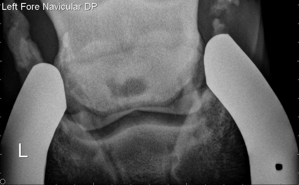
Compared to the right navicular bone (image below), an irregularly shaped lucency (dark circle) is present in the center of the navicular bone (image above).  Although the gelding was not lame, the exam was stopped and the horse was FAILED for sale and intended use.  Everyone involved (including myself) were very surprised with the radiographic findings. However, navicular bone cysts are significant findings and will likely result in poor performance and lameness at some point in the future. As I tell my clients, my crystal ball is "cloudy" at best; however these findings are considered a deal breaker. 


This case represents another example of the importance of a thorough prepurchase exam when acquiring a horse of any price (especially a free horse). In addition, I feel strongly that foot radiographs should be a part of all prepurchase exams due to the essential role the fore feet play in equine soundness. 


Friday, September 21, 2012

Sacro Iliac Injection!


The ultrasound image above is centered over the cranial edge of the pelvic rim. The red line corresponds to the pelvic rim and the green line corresponds to the transverse process of the last lumbar vertebrae.


The picture above shows the "cranial" approach with a long spinal needle for treatment of the sacro-iliac joint (SI). From the exterior of the horse it is very difficult to visualize the SI joint however proper alignment of the spinal needle via ultrasound guidance will result in accurate treatment of the SI joint space. The needle is inserted in front of the pelvis and guided just below the rim of the pelvis (see pic below)


This technique requires ultrasound guidance of a skilled operator. The typical complaint from the rider/trainers include poor performance, no obvious lameness, unwilling to maintain a particular lead, cross-cantering, and painful response to deep pressure over the gluteous musculature. The Sacro iliac joint can be visualized per rectum and may also be evaluated via nuclear scintigraphy. The response to treatment usually requires 10-14 days before a clinical improvement is noted and it can be very significant. The most interesting aspect of SI inflammation is that these horses are rarely lame however their performance as diminished!
The above ultrasound view is per rectum and shows the SI joint. In this case, there are bony changes around the joint which are consistent with arthritis.