PHD Veterinary Service

PHD Veterinary Service

Contact Info

Dr. Porter @ 352-258-3571
portermi.dvm@gmail.com

Read more about Dr. Porter
And PHD Veterinary Services @


Friday, December 13, 2013

Left and Right Laryngeal Hemiplegia in horses

The endoscopic images below are from two different horses with similar clinical complaints yet slightly different physical problems. The gelding in Figure 1 presented for significant respiratory noise during the canter (roaring) and resistance when cantering to the right. The gelding was 12 years old and had a history of "roaring" for multiple years.  In Figure 1, it is evident that there is complete paralysis of the left arytenoid process. Under exercise, the paralyzed process tended to cross over mid-line and prevent adequate passage of air.  He was referred to a surgical facility and a "tie back" procedure was performed. Thirty day post-op endoscopy revealed near complete abduction of the left arytenoid (Figure 2)!! The gelding is significantly improved and will likely perform at a higher level than that prior to surgery.


Figure 1

Figure 2
The endoscopic images in Figure 3 and 4 were from a gelding that presented for significant exercise intolerance at the canter in either direction. The initial endoscopic exam revealed complete paralysis of the right arytenoid process (Figure 3). Paralysis of the right arytenoid is much less common than paralysis of the left arytenoid process and is associated with a worse prognosis. Consultation with an equine surgeon determined that the gelding was NOT a candidate for a "tie back" procedure. In an attempt to improve the gelding's airway, a surgical ablation of several components of the larynx was performed. The thirty day post-op endoscopy revealed a significantly improved airway (Figure 4). Prognosis for this horse as a sport horse remains to be determined however his quality of life has significantly improved! These two cases demonstrate the importance of acquiring the correct diagnosis and surgical intervention via an experienced and highly skilled equine surgeon!! Thank you Dr. John Madison (Ocala Equine Hospital)

Figure 3

Figure 4


Friday, December 6, 2013

Squamous Cell Carcinoma in a Horse

The images below are from 3 different horses with the same disease. Squamous cell carcinoma is a type of cancer that tends to affect the lightly pigmented areas of horses. These areas include the penis (Figures 3-4), sheath (Figures 1-2), muzzle (Figures 5), anus, and the eye lids. Diagnosis should be confirmed via a biopsy and histopathology. This type of cancer does not commonly metastasize to other regions of the body but is locally aggressive. As such, early intervention is the most important component to effectively treating this cancer.

Figure 1


Figure 2
Once the diagnosis of squamous cell carcinoma (SCC) has been confirmed it is my opinion that surgical resection of the tumor should be attempted. This is easier said than done and in some cases surgical removal is not a reasonable option. In some cases, if the lesion is small yet not surgical, cryotherapy with liquid nitrogen can be highly effective. Cryotherapy is recommended for carcinomas near the muzzle and on the penis.  Once the tumor has been removed or if it is not surgical, I recommend proceeding with intra-lesional chemotherapy agents to attack any remaining cancerous cells. It is common to repeat the intra-lesional chemotherapy treatment multiple times.

Figure 3

Figure 4
 It is very important to aggressively treat SCC! The horse in Figure 5 was treated with only surgical resection. Within months of surgery, the tumor returned and invaded the oral cavity. There were no more options for this horse. Horses with SCC need to be treated early and repeatedly until there is no evidence of any remaining cancerous tissue!!
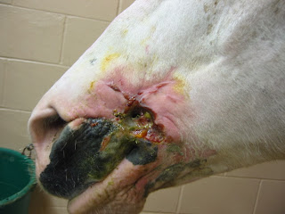
Figure 5