PHD Veterinary Service

PHD Veterinary Service

Contact Info

Dr. Porter @ 352-258-3571
portermi.dvm@gmail.com

Read more about Dr. Porter
And PHD Veterinary Services @


Friday, August 16, 2013

Equine Metabolic Syndrome

The images in Figures 1 and 2 are of an obese donkey. If you follow the dark, dorsal stripe you will notice that the withers of this donkey completely falls to the left side due to the large concenetration of fat that has accumulated within this area!! This condition is also termed "cresty neck".  In Figure 2, the abnormal fat accumulation over the donkey's top line is apparent as is the large deposit of fat just in front of the tail head region! Although slightly humurous, this condition presents a serious medical problem for this donkey. The abnormal accumulation of fat in the area of the neck, top-line, tail head and internally can predispose donkeys and horses to a condition known as metabolic syndrome.

Figure 1

Figure 2
Metabolic syndrome occurs most commonly in over-conditioned horses but has a species predilection for Morgans horses, Arabian horses, Tennessee walking horses, and ponies. The condition is some what similar to adult onset diabetes in humans, which also results in a state of insulin resistance. The serious and some times deadly complication of metabolic syndrome is laminitis!! (Figure 3 and 4). The association between insulin resistance and laminitis is not well understood however in humans with adult onset diabetes, the high circulating levels of glucose are toxic to blood vessels and may cause loss of limb due to poor perfusion. This has not been proven in horses but sounds very similar to what happens in the case of laminitis!  These cases typically present to me AFTER the horse has already foundered and there is a long history (6-24 months) of intermittent lameness, coffin bone rotation, and recurrent foot abscesses!! In my experience, these horses will NOT improve until the insulin resistance is documented and treated!! Unfortunately, if the condition is not diagnosed early enough, severe coffin bone rotation may occur resulting in protrusion of the coffin bone through the bottom of the foot and humane euthanasia (Figure 3 and 4) !


Figure 3


Diagnosis simply involves a blood sample collected from a fasted animal that compares the level of glucose and insulin simultaneously! Occasionally, a glucose challenge may be necessary to establish that a horse is glucose intolerant and insulin resistant. Once diagnosed, the treatment involves regulating the caloric intake of the animal and medical management with thyroid hormone supplement. It is very important to understand that these horses do NOT have a thyroid deficiency!! We are merely increasing the metabolic demand by treating with thyroid hormone supplementation and forcing the animal to lose body fat!!  In addition, to the medical management, the caloric intake is carefully regulated between limited pasture access and low starch diets. Finally, the farrier plays a integral part in helping those horses that have foundered due to the metabolic syndrome. Interestingly, these horses often remain tender-footed regardless of the type of shoe UNTIL the insulin resistance is properly regulated. 

You can read more about equine metabolic syndrome at:


Friday, August 9, 2013

Chronic Obstructive Airway Disease in a Horse

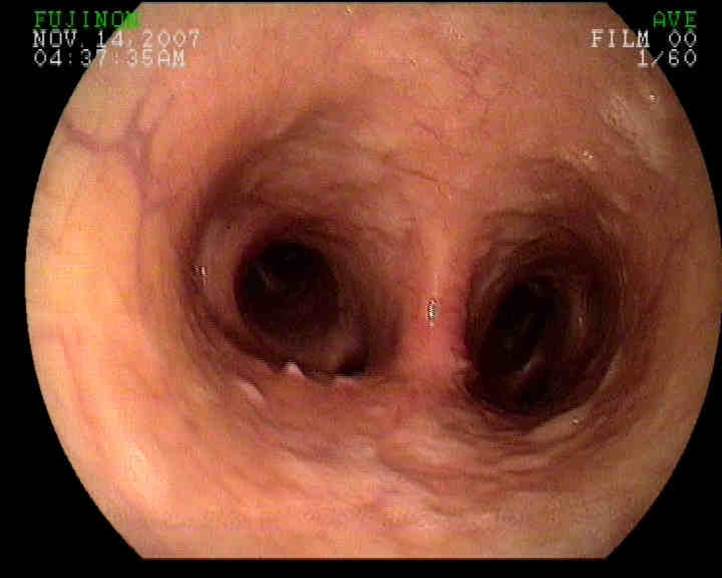
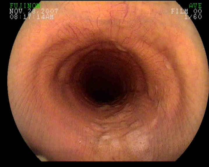
The endoscopic image in Figure 1 is from within a horse's trachea. In Figure 1, the endoscope is positioned just in front of the main bifurcation of the trachea where it splits into the right and left bronchi (Figure 2) . This is just in front of the horse's heart also known as the carina! The purpose of passing an endoscope down to this level of the horse's respiratory tract is to evaluate for the accumulation of debris (feed material, pus and/or blood) and to perform a diagnostic procedure called a bronchoalveolar lavage (BAL). In this particular case, the horse presented for a chronic cough and exercise intolerance.


Figure 1

Figure 2

As the endoscope was passed down the trachea there was a moderate amount of white foamy fluid along the ventral aspect of the trachea (Figure 2 and 3). This debris is consistent with sputum that originates within the lung tissue and is coughed into the trachea. This finding can be consistent with either pneumonia (bacterial infection) or an allergic airway condition that is also known as COPD/Heaves! To determine the cause of the sputum, a BAL is performed. In this procedure, the endoscope is passed into the primary bronchi until it is lodged within the bronchi. At this point a small volume of sterile saline is passed into the bronchi through the scope and then collected via aspiration. The fluid is analyzed to determine the percentage of different cell types and from this data, it can be determined if the horse has an infection or allergic airway disease.

Figure 2

Figure 3

The importance of determining which disease process is causing the sputum is that treatment for allergic airway disease involves systemic corticosteroid administration which will lower the horse's immune system and significantly worsen any bacterial infection the horse may have!! Hence, it is key to perform the BAL  prior to any treatment to determine if the horse needs antibiotics or steroids or maybe antibiotics plus steroids!! The BAL can be performed stall-side with a sedated horse.

Friday, July 26, 2013

Plantar Annular Ligament Desmitis in a Horse


A 10 year old gelding presented for bilateral rear-limb lameness associated with excessive "wind puff" formation. The gelding typically had some wind puff formation above the rear fetlock joints but it had suddenly increased and subsequently the lameness was noted. Physical exam noted moderate to severe fluid accumulation within the distal flexor tendon sheaths, otherwise known as wind puffs (Figures 1-2). The fluid within the flexor tendon sheath tends to accumulate at the top of the sheath due to a very thin band of connective tissue that spans the back or plantar/palmar aspect of the fetlock. This tissue is called the plantar/palmar annular ligament (PAL) of the fetlock (Figure 2 (blue circle))

Figure 1
When fluid begins to accumulate within the tendon sheath due to simple tenosynovitis of the sheath or a tendon/ligament injury, the PAL becomes restrictive resulting in a "wind puff" (Figure 2, red circle), which consists of a pocket of fluid noted just above the fetlock joint. As the fluid increases in volume the PAL is stretched and inflammation develops within the ligament. Once inflammation develops within the PAL, lameness in that limb is common.

Figure 2
 Diagnosis is confirmed via ultrasonic exam. In Figures 3-4, the flexor tendons within the sheath are imaged and include the superficial flexor (SDF: blue) and deep digital flexor tendon (DDF: green). In addition, the PAL is visualized directly above the SDF tendon. Normally, the PAL is very thin and only measures 2-4mm in thickness. In this horse, the PAL measured nearly 10mm in thickness (blue arrows) and there were pockets of edema (black areas) along with poor fiber alignment! Chronic thickening of the PAL ligament may result in the inflammation within the SDF tendon which will significantly worsen the prognosis and lameness!  For this reason, careful examination via ultrasound is key.

 (Figure 3
 In this particular horse there does not appear to be involvement of the SDF or DDF tendons. The PAL appears very thick and inflamed. In Figure 4, the same area is imaged in a cross sectional view (left image) and a transverse view (right image). The significant amount of edema and disrupted fiber pattern within the PAL is quite evident in the both views!! Management of this condition involves corrective shoeing, topical anti-inflammatory application, and in my opinion surgical intervention. Once the PAL is grossly thickened, it is my experience that only surgical transection of the PAL ligament will provide a long term solution for the horse. It is very important to thoroughly ultrasound the limb through the pastern area to ensure that no ligaments or tendons are involved!! Finally, wind puffs are a common occurrence in many sport horses and usually indicate that the horse is in steady work. However, asymmetric wind puffs or a sudden increase in size is not normal and merits examination by your veterinarian!!

Figure 4

Friday, July 19, 2013

Thin Soles in a Horse!!


The radiographs below in Figures 1-3 belong to a middle-aged QH gelding that presented for severe lameness. The gelding was recently trimmed by the farrier and had a history of mild to moderate foot pain.
On presentation, the gelding was very sore when asked to walk on a firm surface and was not willing to walk in a tight circle! There were NO pounding digital pulses however firm pressure, via my finger tips, over the sole region resulted in a strong pain reaction by the gelding. Radiographs were taken and severely thin soles were diagnosed in both front feet (Figure 1 and 2). The actual thickness of the sole was difficult to measure due to the thinness but was approximated at 2-3mm (Figure 3). Less than 5mm of thickness essentially corresponds to 1 swipe with the hoof knife!!

Figure 1

Figure 2


Figure 3


In Figure 4, there is a radiograph of a normal horse's foot. The sole depth at the toe region is approximately 15-20mm. In my experience, when the sole thickness is less than 10mm, there is increased likelihood of foot pain from concussion with the ground. Thin soled horses are at high risk for sole bruising and sub-solar abscess development. Unfortunately, some horses are "naturally" thin soled due to poor genetics and MUST be managed with shoes. The corrective shoeing is simple and may include a rim pad, leather pad , or soft pour-in pad. I strongly recommend radiographic evaluation PRIOR to any foot trimming to determine the sole thickness of horses that have a history of chronic foot pain!

Figure 4

Friday, July 12, 2013

Sand Impaction in a Horse

A six year old gelding presented for recurrent low grade fever and intermittent diarrhea. At the time of the initial exam the gelding was alert and showing no signs of discomfort. Vitals were normal and body temperature was 100.5 degrees.  A blood sample was collected for complete blood count (CBC) analysis and a rectal exam was performed. A large, firm structure was palpated along the left flank of the horse which was consistent with an impaction of the "pelvic flexure".  The fecal material removed from the rectum was partially formed and had a grainy feel to it. Several hand fulls of manure were placed in a rectal sleeve and the sleeve was filled with water (Figure 1).  A significant amount of sand was identified in the sleeve filled with water!

Figure 1

 The gelding was treated aggressively with water and mineral oil via a naso-gastric tube however; his condition worsened and he was referred to a surgical facility. Despite knowing that the gelding had a pelvic flexure impaction which is usually managed medically, the gelding was taken to surgery due to refractory pain. Abdominal exploration revealed an extremely large, firm impaction of the left colon which "bottle necked" at the pelvic flexure (blue arrow in Figure 2).  Discovery of such an impaction during surgery typically results in making a small opening in the colon wall and resolving the impaction. However, a small tear was discovered along the edge of the impaction which had resulted in fecal contamination of the abdomen. Prognosis was poor and the owners opted for humane euthanasia.

Figure 2
This case of sand impaction in a horse is sadly NOT uncommon in the state of Florida! The accumulation of sand within the colon of a horse is typically a result of poor pasture conditions and can develop very quickly. There are several "hair-pin" turns within the large colon of horses which create ideal conditions for the accumulation of sand and other debris. The pelvic flexure is one of those "hair-pin" turns and is one of the most common locations for colon impactions in horses. Unfortunately, once the sand impaction develops it is very difficult to resolve by providing oral medication such as psyllium (Figure 3)  and mineral oil (Figure 4).

Figure 3
Figure 4
The clinical indicators of sand accumulation in horses include intermittent diarrhea, low white blood cell count, fever and plus/minus colic. The sand is abrasive to the lining of the colon which results in diarrhea and sequestration of white blood cells in the colon wall. Horses with sand accumulation within their colons often have a week or month-long history of diarrhea! This is the time to treat the horse with sand clearing products. Once the horse becomes completely obstructed, surgery may be the only option to resolve the impaction. There are several products available to treat horses on a monthly basis as "sand clearing" products however once sand has accumulated within the colon, these products are not effective. They are more effective if used as products to prevent sand accumulation in combination with good pasture management! This condition is treatable but MUST be addressed when clinical signs first develop!!

Friday, July 5, 2013

Sub-solar Abscess in a Horse!!

Figure 1

The radiograph in Figure 1 is of a horse's hind foot. This gelding has been suffering from chronic laminitis/founder for over 1 year. When I first examined this horse, there was nearly zero sole depth between the coffin bone and the bottom of the foot. In addition, the coffin bone was reduced in size due to gradual degradation (pedal osteitis) of the coffin bone. The pedal osteitis developed due to the chronic pedal bone rotation that is evident in Figure 2 (non-parallel red lines). It was decided that a deep digital flexor tenotomy combined with appropriate shoeing may help stabilize the foot and promote sole growth. Over 4-6 months after the surgery, the gelding's sole depth increased significantly, indicating normal hoof growth and he was more comfortable on this limb.

Figure 2

Recently, the gelding's lameness worsened and he was non-weight bearing when examined. A lateral radiograph was performed (Figure 1-3). A large, radiolucent area (red circle) was noted in the toe region of the foot with a track that appeared to be traveling toward the coronary band (Figure 3). These findings are consistent with a large, sub-solar abscess! 

Figure 3
The shoe was removed and a large "hole" was noted at the apex of the frog that extended into a cavity with the circumference of a golf ball! The abscess appeared to be draining however it involved a significant amount of space between the bottom of the sole and the coffin bone. After removing the shoe, the foot was soaked in a warm epsom salt solution and then was "packed" with a strong disinfectant. The gelding's lameness improved within 24 hrs however he remains lame at the walk. His return to soundness will take some time and most likely require a specialized shoe known as a hospital plate. Recurrent foot abscess formation is a common problem among chronically foundered horses and highlights the importance of regular trimming/shoeing plus radiographic examination!

Figure 4


Friday, June 28, 2013

Habronemiasis (Summer Sore) in a Horse!


The images in Figure 1-3 are from a horse that presented for a large, painful swelling along the mare's ventral abdomen. The mare had been treated by the referring veterinarian with anti-inflammatory medication and oral antibiotics with minimal improvement. A core biopsy revealed chronic inflammation with no specific cause identified and an ultrasound exam merely revealed diffuse edema. Close examination of the swelling (Figures 2-3) noted yellow granules which were consistent with sulfur granules. This finding is consistent with 2 diseases in horses which are cutaneous pythiosis and cutaneous habronemiasis. Identifying which pathogen is causing the lesions can be tricky and involves skin biopsies with special staining for pythiosis.


Figure 1

Figure 2

Figure 3

According to the Merck Veterinary Manual, "cutaneous habronemiasis or summer sores are a result of fly larvae which enter existing wounds or moist skin and migrate into the tissue causing irritation and a chronic granulomatous reaction".  As such, management of this condition involves fly control, medication for encysted larvae, AND medication for inflammatory reaction. Fly control is an ongoing issue at most farms and becomes increasingly important as the warm, summer months approach. The most common method for treating for the encysted larvae is a simple dose of oral ivermectin. Unfortunately, this is were most horse owners stop with treating summer sores. There are a multitude of topical therapies which include varying amounts of ivermectin and antibiotics. Unfortunately, these topical therapies are NOT appropriate for treating the chronic inflammatory reaction. As a result, many summer sores continue to increase in size and result in horrible wounds as imaged in Figures 4 and 5. These represent the MOST common location for summer sores in horses that being the lower limbs.

Figure 5

Figure 4
The large swollen plaque across the ventral abdomen is a uncommon location for a summer sore as is the upper lip noted in Figure 5. However, these lesions can develop on any part of the horse's body that is accessible by a fly!! It is critical that the horse owner understands that the chronic, inflammatory reaction is a result of a hypersensitivity or allergic reaction to the encysted larvae, before and AFTER it has been effectively killed with systemic ivermectin. As such, the horse must be treated for the hypersensitivity with products that contain immune suppressive doses of corticosteroids. In small, focal cases, the lesions are effectively treated with topical treatment however large summer sores will need systemic corticosteroid administration!! In my experience, this is the ONLY way to effectively rid the horse of this condition. Unfortunately, once a horse develops summer sores, they will be predisposed the following fly season to re-develop sores in the same regions. I have developed a highly effective topical cream with the help of a pharmacy and would be happy to dispense this product to owners of horses that are dealing with this highly annoying condition!!

Figure 6

Thursday, June 20, 2013

Paranasal Sinus Cyst in a Horse

Figure 1A
Figure 1B


The radiographs in Figure 1-3 correspond to a horse that presented for chronic nasal discharge. The discharge was bilateral and there was a reduction in the air flow through the nasal passages. Due to the reduced airflow, it was decided to first radiograph the gelding's skull and then potentially scope the upper airway.


Figure 2A
Figure 2B


Radiographic exam revealed a soft tissue mass that was located in the maxillary sinus. In Figures 1-2 A and B the soft tissue mass appears as a round, white structure that is highlighted by the red dotted circles. In Figure 3 there appears to be a soft tissue mass in both the left and right maxillary sinus (red dotted circles). The mass appears larger on the left side of the skull.
The gelding was NOT sedated for endoscopic exam in order to reduce the likelihood of respiratory distress.

Figure 3
 In Figure 4, an endoscopic image reveals a smooth, soft tissue tumor/mass that is obstructing the nasal passage. Moderate amounts of purulent debris can be seen accumulating around the soft tissue mass. Based on the endoscopic and radiographic findings, the gelding was referred for surgical exploration. At surgery, a large paranasal cyst was exposed in the left maxillary sinus and a small cyst was identified in the right maxillary sinus. Both cysts were removed and the gelding recovered uneventfully. It is likely that the cysts began to develop early in life for this gelding and did not become clinically relevant until they grew into the nasal passages. Once they are removed via surgical intervention the prognosis is very good !!


Figure 4


Friday, June 14, 2013

Shoulder swelling in a Horse

The mare in Figure 1 presented for a complaint of sudden onset severe swelling at the point of the right shoulder and near non-weight bearing lameness of the same limb. The mare is normally kept with other horses and there is no history of a "equine dispute" or evidence of external trauma. On presentation, the swelling consists of significant edema and the mare is considerably "tender" to pressure and palpation of the swelling. Manipulation of the limb was limited due to the significant pain response that resulted. Radiographic exam of the horse's shoulder is a viable option however due to the pain on manipulation and significant soft tissue swelling, an ultrasound exam was more highly indicated.

Figure 1
The images in Figure 2 and 3 correspond to the soft tissue swelling noted in Figure 1. A large, fluid filled pocket was noted deep to the skin surface. The fluid had a mixed pattern of echogenicity suggesting that the fluid consisted of cellular debris mixed with blood/serum. These findings are most consistent with a large subcutaneous abscess. The two most likely causes for abscess formation in this area would be trauma induced or abscess formation from a bacteria called corynebacterium pseudotuberculosis. This condition is also known as "Pigeon's fever". Pigeon's fever is a serious condition due to the highly infectious nature of the bacteria AND the fact that once the bacteria is present on the premises, it is very difficult to remove from the environment. Correct diagnosis depends on collecting a sample of fluid from the abscess and submitting for laboratory evaluation. Treatment of either condition requires establishing drainage of the abscess and daily  lavage for 10-14 days after drainage. Microbiological identification in this case is pending and the horse is in quarantine on the farm! The abscess was opened and drainage established.

Figure 2


Figure 3
Interestingly, within 7 days of examining the mare above, a gelding presented for similar symptoms. He was found non-weight bearing lame in the pasture with severe swelling of the shoulder region. The referring veterinarian provided digital radiographs which did not identify the cause of the lameness/swelling. On ultrasound exam, a bright, bone-like structure (red dotted line) was noted extending into a fluid filled mass that appeared to be a hematoma (Figure 4 and 5). The bone-like structure was the humerus bone (red dotted line) and it was fractured plus displaced. This gelding was humanely  immediately euthanized!

Figure 4

Figure 5
Although these cases are quite different in outcome, they demonstrate the benefit of soft tissue ultrasound with regards to assessing "swellings". As demonstrated it is possible to evaluate soft tissue changes as well as bony changes.


Friday, June 7, 2013

Superficial digital Flexor Tendonitis (Bowed tendon) in a Horse!

A  twenty year-old mare presented for sudden, severe swelling of the left front lower limb. The mare was nearly non-weight bearing lame and had been seen galloping around her paddock moments before. Prior to my exam, the mare was stalled for 7 days and treated with ice and systemic non-steroidal mediation (phenylbutazone). On presentation there was a noticeable "profile" to the lower portion of the forelimb (Red line:Figure 1). The mare was moderately positive to pressure over this swelling and was lame at the walk. On palpation, there was no distinction between the superficial digital flexor tendon and the deep digital flexor tendon.
Figure 1
Ultrasound examination of the lower limb, beginning just below the knee (carpus) revealed inflammation of the superficial digital flexor tendon (SDF). The inflammation was moderate just below the knee (Figure 2) however became severe further down the leg (Figure 5). In Figures 2 and 3, moderate to severe edema (dark tissue) is noted with pockets of disrupted fiber pattern. The image in Figure 4 is of the opposite limb for comparison to /the abnormal limb. In Figure 5, the SDF tendon (Red arrows) is severely enlarged and there is no apparent pattern with regards to the fiber pattern. These findings are consistent with a near rupture of the SDF tendon.

Figure 2

Figure 3

Figure 4

Figure 5
Although the outward and inward findings are severe, the prognosis for pasture soundness is good. It will take 6-12 months for this type of injury to heal with a fibrotic scar but the mare is likely to return to soundness as a pasture horse. Unfortunately, her prognosis as a riding horse is poor and it is not recommended to return this mare to any type of forced work due to her age and degree of injury. Tendonitis of the superficial flexor tendon can occur in various regions of the lower limb. The closer to the knee/carpus (High bow), the worst the prognosis, even for pasture soundness. This is due to the constant irritation of the tendon when the horse advances the limb. In addition, horses that suffer a "bowed" SDF tendon are at increased risk for another injury to the same tendon and it commonly occurs above or below the original injury. My recommendation for rehabilitative care includes the following:

1: Limited activity: preferred stall rest with hand walking for 2 months followed by small paddock turn-out
2: Daily ice therapy for 2-3 weeks
3: Topical non-steroidal cream: Surpass
4: Consider Extra-corporeal shockwave treatment and PRP injection.