PHD Veterinary Service

PHD Veterinary Service

Contact Info

Dr. Porter @ 352-258-3571
portermi.dvm@gmail.com

Read more about Dr. Porter
And PHD Veterinary Services @


Friday, July 27, 2012

Equine Pharyngitis!!


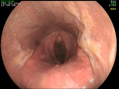
The image above was from a 10 year old gelding that presented for a history of coughing and mild nasal discharge. The cough was intermittent and would vary from day to day. Recently, the cough had worsened as had the summer heat! Based on the history, the most likely cause was predicted to be heaves (AKA: COPD). However, when the endoscope was introduced into the gelding's upper airway it was clear that we were dealing with something quite different. There was evidence of pharyngeal scarring (cicatrix) and active pharyngeal ulceration  in a 360 degree (red circle below) range.  These findings were consistent with active inflammation and chronic changes. In addition, there was chronic, active inflammation of the arytenoids (green arrow).


The cause of pharyngeal inflammation/ulceration varies and is often never determined. In some cases, it may be complicated by bacterial infection. In some cases, the inciting cause is presumed to be an environmental irritant that the horse is chronically exposed to. The chronic inflammation results in scarring or cicatrix formation that can result in narrowing of the pharynx. Pharyngeal cicatrix formation is a serious complication that has no reliable remedy!  As such, a chronic couch (>2 weeks) should be evaluated via endoscopy prior to the development of scarring. Pharyngeal inflammation is typically treated with forced rest and topical antibiotics/steroids in the form of a nasal flush. 


Friday, July 20, 2012

Flood = Mosquito = Sleeping sickness (AKA: Encephalitis)

Yearling paint with EEE (non-vaccinate)
The gelding died within 2 days of showing clinical signs!

This year has been a record year for many parts of Florida with regards to rain fall. Although this has been a great thing for our over-all drought conditions, it has created a situation that may result in the awakening of a sleeping monster, also known as equine encephalitis. Depending on the virus, horses in the southeastern United States may be afflicted with the virus that causes Eastern Equine Encephalitis (EEE) or West Nile Encephalitis. Sadly, the mortality rate for EEE in non-vaccinate horses is nearly 100% and approximately 30% in non-vaccinate horses with West Nile virus. These viruses are transmitted via the mosquito and tend to be most prevalent during the warmer months of the year. Due to the excessive rain in Florida, there are many areas with various amounts of flooding and sitting water. These are ideal conditions for an explosion in the mosquito population!  Already in Alachua county, a sentinel chicken tested positive for the virus that causes EEE.


The good news is that these diseases are nearly 100% preventable through proper vaccination. At a minimum, horses should be vaccinated 2x per year for these dreaded diseases. However, with the explosion of the mosquito population this year, I recommend vaccinating 3x per year and vaccinating your horse within the the next 30 days if it has been more than 3 months since the their last vaccination. 
Please contact your veterinarian today and make plans to protect your horses!! 


You can read more about these conditions at the following site

http://www.aaep.org/eee_wee.htm


Friday, July 13, 2012

Cleft Palate!

Image 1
The first two images are from a yearling thoroughbred that presented for a long history of nasal discharge, coughing while eating, and the presence of water exiting from nostrils while drinking. The filly had been treated with antibiotics prior to the exam. Endoscopic exam revealed a defect within the soft palate that extended nearly the entire length of the soft palate. In addition, the epiglottis was entrapped by the aryepigltottic fold. 

Image 2
 The defect within the soft palate is known as a "cleft palate" and most likely has been there since birth. Fortunately, this is a relative rare condition! Normally, the palate plays the role of separating the nasal passages from the oral passage. Hence, a horse is an obligate nasal breather and the palate keeps water and feed from entering the airway. When a cleft palate exists, there is communication between the air passage and oral passage resulting in feed and water going the "wrong way". This may consist of feed material exiting the nostril and/or traveling down the trachea into the lungs. If these horse go undiagnosed they are typically "poor doers" with failure to thrive secondary to chronic respiratory infections. Sadly, there is little to be done to help such a severe cleft palate. Less severe cases may be addressed with surgical intervention with mixed results. 


Image 3

The third image if that of a "normal" horse. The epiglottis is visible and is resting on top of the complete soft palate. Diagnosis of a cleft palate is often made immediately after birth during a thorough post-foaling exam. The veterinarian should palpate the length of the hard and soft palate with their finger to make sure it is normal. If missed at foaling, the diagnosis can be made quite simply with an endoscopic exam. 


Friday, July 6, 2012

Friday, June 22, 2012

Equine Hirsutism


The horses pictured below are both suffering from a condition known as equine hirsutism. The condition can be described as an irregular or excessive hair coat throughout the year. Normally, a horse will develop a winter coat for the cooler months and then shed the coat throughout the spring months. Horses suffering from  hirsutism will develop a thick coat throughout the year and will require regular, full body clips to keep cool during the hot summer months. 


By far, the most likely cause of equine hirsutism is equine cushing's disease. This is an endocrine disorder that is centered in the brain of the horse and results in excessive cortisone production. Much more serious than a persistent shaggy hair coat, horses with cushing's disease are highly susceptible to founder (laminitis) and a compromised immune system. Testing for equine cushing's disease is a tricky process and may involve measuring serum levels of cortisol and ACTH. In addition, horses can be tested via a Dexamethasone suppression test which may be risky procedure if the horse has a history of founder. As such, it is commonly recommended that if the horse is suffering from hirsutism, it should be assumed that the horse is likely to be also suffering from equine cushing's disease and should be treated for the latter. Recently, the preferred drug of treatment knows as Pergolide became available to equine owners in the form of a FDA approved drug called Prascend. Horses as young as 10 years of age can develop these symptoms and should be treated sooner than later!


Read more about equine cushing's disease @


Friday, June 15, 2012

When Is A "Wind Puff" Abnormal?

Figure 1
 The "wind puff" noted in the right hind leg is not necessarily an immediate cause for alarm (Figure 1) . However, when you compare it to the left hind leg, there is a significant difference in the size of the wind puff between legs (Figure 2). This is a reason for concern!

A "wind puff" refers to the accumulation of synovial fluid with the distal flexor tendon sheath of the lower leg. All 4 limbs have a distal flexor tendon sheath and they extend from just above the fetlock down to the coffin bone area. Most people notice the swelling once it is above the fetlock joint however there is often significant fluid within the pastern area that goes unnoticed. For many horses that are in regular work a mild amount of fluid accumulation within the flexor tendon sheath is normal and should be symmetrical. When there is more fluid in one leg or a sudden increase in fluid then there is reason to be concerned.

Figure 2

Within the flexor tendon sheath there are a various tendons and ligaments. Any inflammation within these tendons and ligaments may result in a sudden increase in fluid within the sheath and eventually a reduction in performance. To thoroughly assess these soft tissues, an ultrasound exam should be performed and the soft tissue structures above AND below the fetlock should be evaluated. The image below is that of a abnormal deep digital flexor tendon. The area of concern is at the level of the fetlock joint and within the flexor tendon sheath.  The tendinitis has been chronic and there is evidence of tendon mineralization!. This horse was lame on presentation and there was significant fluid within the distal flexor tendon sheath.


Figure 3


Friday, June 8, 2012

Clostridial Myositis and Banamine Injections

Intra-muscular Banamine is not recommended by this veterinarian!


The picture above is that of a horse that was treated by its owner with 10cc of Banamine (Flunixin Meglumine) in the muscle (IM) of the left hind leg for colic symptoms. The good news was that the horse's symptoms of colic resolved however 2 days later the injection site was swollen and painful to the touch. The following day, the horse was febrile and lame at the walk. The wound was examined via ultra-sound and a large, fluid filled pocket was noted deep to the swelling. The pocket was drained of purulent (pus) fluid and the horse was started on antibiotics. Fortunately, the horse recovered fully with only a minor scar.

This type of reaction may occur secondary to any intra-muscular injection. However, the big concern with injection site abscesses caused by IM Banamine is the association with the bacteria Clostridium and the resulting Clostridial myositis.  These bacteria are anaerobic and thrive in low oxygen environment, hence treatment involves aggressive surgical exposure and drainage of the contaminated tissue. Severe Clostridial myositis can readily result in death of the horse!!

The preferred route of administration for Banamine is either intra-venous or by mouth.

Friday, June 1, 2012

Core Biopsy Please!!

The only way to know for sure would require a core biopsy. This procedure is quite simple to perform in the field and will save significant time with regards to treatment and resolution of clinical signs. The first image was treated unsuccessfully as a "summer sore" with medication that actually worsened the clinical signs. The biopsy results were consistent with Pythiosis and aggressive medial/surgical treatment was instituted. The prognosis for full recovery is not good once there has been significant disruption of the soft tissue structures and in a case that involves the lower pastern/coronary band area. Early diagnosis is key!


The case below was treated as a suspect "summer sore" with minimal improvement. Biopsy results indicated that it in fact it was a case of Habronemiasis that was not responsive to the treatment provided (systemic and topical ivermectin). Habronemiasis or "Summer Sores" is a condition that results from flies depositing larvae within the skin causing a significant allergic reaction. Treatment involves medication to kill any living fly larvae and most importantly medication to suppress the local allergic reaction. Treating with ivermectin systemically and topically is often not enough!! Yet that is how many try to treat this condition! For small lesions, topical treatment with concentrated cortisone can be effective, however, for more aggressive lesions, systemic administration with corticosteroids is necessary. Importantly, systemic treatment with corticosteroids is the treatment you definitely want to avoid if you are dealing with Pythiosis! Hence the importance of a core biopsy.


Friday, May 18, 2012

Equine Strangles

Equine Strangles!!

(Figure 1)


(Figure 2)


The disease process that may cause throat latch swelling AND limb swelling is commonly known as Equine Strangles. This disease is caused by a bacteria known as Streptococcus Equi subspecies Equi. Most commonly it results in enlargement of the lymphnodes within the throat latch area that may narrow the airway of the horse, hence the term "strangles". In severe cases, a temporary tracheotomy is necessary to save the horses life! (Figure 1). These lymph nodes may rupture and drain purulent debris from the outside or from within the guttural pouches (Figure 2). These horses are typically febrile, depressed, and highly contagious! Occasionally, the horse will develop a hyper-immune response known as purpura hemorrhagica. This immune response to the bacteria results in severe swelling of one or multiple limbs (Figure 3)  and can be triggered by the vaccination of a horse that was recently sick with the disease.


(Figure 4)

For more information check out the following links:


Friday, May 11, 2012

Mesotherapy 


These small bumps were caused by micro-injections of medication just under the skin, or also known as Mesotherapy. This treatment has been used effectively for over 30 years in humans and horses. The primary objective of this treatment in horses is to block or break the pain cycle associated with chronic back pain. 

Technically, mesotherapy treatment stimulates the mesoderm or middle layer of skin by stimulating the giant fibers present within this layer of skin. Commonly, a combination of lidocain and dexamethasone is injected via multiple extra small needles (30g). These micro-injections result in stimulation of the giant nerve fibers located within this layer of skin that also communicate with major nerve tracts within the spinal chord. These nerve fibers INHIBIT the conduction of pain signals to the brain.

Typically, one or two mesotherapy treatments are applied to a horse's back and often are combined with shockwave treatments. Mesotherapy is used primarly as an aid to alleviate back pain followed by treatment of the primary cause of the back pain/spasm. This typically involves a source of lameness in either the forelimbs or hindlimbs. In addition, osteo-arthritis of the thoraco-lumbar spine and "kissing spines" are causes of chronic back pain. 

The response to mesotherapy is usually within 2-3 days and may last for 3-4 months.