PHD Veterinary Service

PHD Veterinary Service

Contact Info

Dr. Porter @ 352-258-3571
portermi.dvm@gmail.com

Read more about Dr. Porter
And PHD Veterinary Services @


Showing posts with label michael porter. Show all posts

Friday, September 27, 2013

Hind Limb Proximal Suspensory Ligament Desmitis in a Horse

A 15 year-old mare presented for a 3 week history of rear-limb lameness that was associated with a "drop" of the rear fetlock joint. On presentation there was moderate swelling of the lower limb, just below the hock joint and the mare was lame at the walk. In addition, there was a 90 degree drop of the fetlock/pastern axis as noted in Figure 1.

Figure 1

The primary mechanism involved in "suspending" the fetlock joint and maintaining the proper fetlock/pastern axis is the suspensory ligament (Figure 2). The suspensory ligament originates just below the hock (red arrow) and initially is one structure (body) that travels down the back of the lower limb (yellow arrow). Approximately half way down the canon bone the suspensory ligament splits into a medial (inside) and lateral (outside) branch. The suspensory branches attach to the sesamoid bones which are located just behind and below the fetlock joint. As such, the suspensory ligament helps "suspend" the fetlock joint and a  proper fetlock/pastern axis.

Figure 2
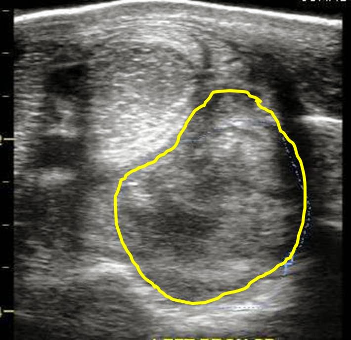
An ultrasound exam was performed to evaluate the entire suspensory ligament. The origin or proximal suspensory ligament is imaged in cross-section in Figures 3-6. The proximal suspensory ligament of the affected limb is grossly enlarged (yellow circle) and the fiber pattern is a mixed pattern with significant edema and evidence of active inflammation! There is a black and grey swirl pattern noted in the proximal suspensory ligament (tissue inside the yellow circle) of the affected limb which is indicative of severe changes.


Figure 3


Figure 4

 When compared to the normal limb, the significant increase in the size of the proximal suspensory ligament is evident. In this case the affected suspensory ligament was 2x the "normal" size. These ultrasound findings confirm the diagnosis of proximal suspensory desmitis of the hind limb. The prognosis for this injury is poor for return to riding and guarded for return to pasture soundness. Once the fetlock has "dropped" the physical changes to the suspensory ligament CANNOT be reversed!! Treatment is aimed at slowing the progress of the condition and attempting to provide pain relief to the horse. In my experience, corrective shoeing is the MOST important aspect of managing this condition.

Figure 5
  
Figure 6
A "fish tail" bar shoe is strongly recommended for this condition. The shoe should be set extra full such that approximately 1.5 to 2 inches of shoe extended BEHIND the heel bulb. Any kind of a wedge is CONTRAINDICATED in this condition! In addition, daily treatment with ice packs over the proximal suspensory ligament followed by topical Surpass cream are indicated to reduce inflammation and provide pain relief. With corrective shoeing, adequate pain relief, and supervised turn-out, these horses may return to pasture soundness however such a condition carries a guarded prognosis.

Figure 7


Friday, June 7, 2013

Superficial digital Flexor Tendonitis (Bowed tendon) in a Horse!

A  twenty year-old mare presented for sudden, severe swelling of the left front lower limb. The mare was nearly non-weight bearing lame and had been seen galloping around her paddock moments before. Prior to my exam, the mare was stalled for 7 days and treated with ice and systemic non-steroidal mediation (phenylbutazone). On presentation there was a noticeable "profile" to the lower portion of the forelimb (Red line:Figure 1). The mare was moderately positive to pressure over this swelling and was lame at the walk. On palpation, there was no distinction between the superficial digital flexor tendon and the deep digital flexor tendon.
Figure 1
Ultrasound examination of the lower limb, beginning just below the knee (carpus) revealed inflammation of the superficial digital flexor tendon (SDF). The inflammation was moderate just below the knee (Figure 2) however became severe further down the leg (Figure 5). In Figures 2 and 3, moderate to severe edema (dark tissue) is noted with pockets of disrupted fiber pattern. The image in Figure 4 is of the opposite limb for comparison to /the abnormal limb. In Figure 5, the SDF tendon (Red arrows) is severely enlarged and there is no apparent pattern with regards to the fiber pattern. These findings are consistent with a near rupture of the SDF tendon.

Figure 2

Figure 3

Figure 4

Figure 5
Although the outward and inward findings are severe, the prognosis for pasture soundness is good. It will take 6-12 months for this type of injury to heal with a fibrotic scar but the mare is likely to return to soundness as a pasture horse. Unfortunately, her prognosis as a riding horse is poor and it is not recommended to return this mare to any type of forced work due to her age and degree of injury. Tendonitis of the superficial flexor tendon can occur in various regions of the lower limb. The closer to the knee/carpus (High bow), the worst the prognosis, even for pasture soundness. This is due to the constant irritation of the tendon when the horse advances the limb. In addition, horses that suffer a "bowed" SDF tendon are at increased risk for another injury to the same tendon and it commonly occurs above or below the original injury. My recommendation for rehabilitative care includes the following:

1: Limited activity: preferred stall rest with hand walking for 2 months followed by small paddock turn-out
2: Daily ice therapy for 2-3 weeks
3: Topical non-steroidal cream: Surpass
4: Consider Extra-corporeal shockwave treatment and PRP injection.

Friday, May 24, 2013

Testicular Enlargement in a Stallion

A 19 year-old stallion presented for the complaint of testicular enlargement. On physical exam, the left testes palpated firm, non-painful and was approximately 30-40% larger than the right testes (Figure 1). The potential differentials for an enlarged testes in a stallion include testicular neoplasia (cancer), hydrocele, scrotal hernia, testicular torsion, and orchitis. Definite diagnosis is based on ultrasound exam and histologic exam of the abnormal tissue. Ultrasound exam of the stallion's testes was performed during the initial visit.

Figure 1
In Figures 2 and 3, the left testes is imaged via ultrasound. The testicle appears to be divided by two tissues-types which vary in density or echogenicity (degrees of brightness). The normal tissue appears brighter or hyper-echogenic whereas the abnormal tissue appears darker or less echogenic. The edges of the abnormal tissue are distinct and there are multiple, small "stars" or areas of increased echogenicity throughout the abnormal tissue. 


Figure 2

Figure 3
Ultrasound exam of the right testes revealed a homogeneous tissue consistent with normal testicular structure (Figure 4). The echogenicity did NOT vary throughout the testicle and the over-all size of the testicle was less than the left testicle. In Figure 5, the left and right testicles were imaged simultaneously and the difference in testicular architecture becomes more clear when comparing the "normal" right tests to the "abnormal" left testes. 

Figure 4


Figure 5

Testicular neoplasia or cancer is relatively rare in stallions compared to dogs. The types of cancer include seminomas, sertoli cell tumors, and leydig cell tumors. Histopathologic exam is required to determine the type of testicular cancer. All three types of cancer will result in compromise of normal sperm-producing tissue and thus reduction in fertility. Seminomas are the most common of the three types of testicular cancer in stallions and may have a high degree of malignancy and invasiveness. Scrotal hernia, testicular torsion and orchitis are additional causes of testicular enlargement which are typically associated with varying degrees of pain and inflammation. Ultrasound findings of these conditions may include small intestinal loops, dilated vascular supply and pockets of purulent debris. Similar to testicular cancer, these conditions typically alter the conditions for normal sperm production and will reduce fertility. A more benign cause of testicular enlargement is known as a hydrocele. This corresponds to an accumulation of abdominal fluid within the scrotal sac. Although benign, the increase in fluid may cause change in temperature and pressure which will also affect sperm production. This condition is more easily confirmed with ultrasound exam alone!.

The stallion in this case is scheduled for surgical removal of the affected testes for definitive identification of the cause for the testicular enlargement. Stay tuned!! 


Friday, May 17, 2013

Injectable Supplements for Horses

Listed below are the most common (non-steroidal) injectable supplements administered to horses to treat for active arthritis and hopefully slow down the development of arthritis. These medications vary in cost, method of administration, and approved use. One of the best known and most commonly used is Adequan. The active ingredient in Adequan is polysulfated glucosaminoglycan (PSGAG) and is FDA approved for intra-muscular and intra-articular use in horses. It is reported that PSGAG-containing products, such as Adequan, work by reducing inflammatory mediators within the joint and help maintain healthy cartilage. Adequan may be given in a loading dose initially for 5-7 treatments and then once monthly or may be administered every 6 months as a loading dose of 7 treatments that are 4-7 days apart.
Figure 1: Molecular structure of Adequan

Figure 2: Intra-articular Adequan

Similar to Adequan , a relatively new supplement in the US is known as Pentosan. The active ingredient in Pentosan is Pentosan Polysulfate Sodium (PPS) and is distinct in molecular structure from Adequan (Figure 1 versus Figure 3) . In humans, PPS is used to treat bladder pain associated with cystitis. In addition, clinical research in humans suggests that PPS may reduce clinical signs associated with arthritis. In horses, clinical and empirical data indicates that regular administration of PPS-containing products reduces clinical signs associated with osteoarthritis. Pentosan is available in the United States as a generic product that is sold through compounding labs (Figure 4). In my practice, it is commonly administered as an intramuscular injection, initially as a loading dose (5 injections, 1 week apart) and then 1-2x per month.

Figure 3: Molecular structure of PPS


Figure 4: Generic Pentosan

A PPS containing product that is licensed for use in horses in the US and FDA approved is PentAssuie (Figure 5) . This product  contains both Pentosan AND N-acetyl glucosamine (PSGAG). N-acetyl glucosamine is a PSGAG but is slightly different in structure compared to Adequan. PentAussie is approved for post surgical lavage and is considered "off label" when administered intra-muscularly. 

Figure 5
The product known as Legend is an injectable supplement approved for use in horses that contains Hyaluronate sodium which is non-sulfated glycosaminoglycan. Hyaluronate is a normal constituent of joint fluid and plays a key role in regulating inflammation within the joint. The product Legend is typically administered intravenously however products containing hyaluronic acid are injected directly into the joint. The frequency of IV administration varies from prior to competition to monthly.  

Figure 6

 Polyglycan (Figure 7) is an injectable supplement that contains a mixture of several productions (Figure 8). It is approved for use in horses but NOT for intravenous administration, which is the most common route of treatment! The product contains hyaluronate, acetyl glucosamine, and chondroitin sulfate (active ingredient in Cosequin).  In my practice, this supplement is administered several days before competition and/or on a monthly basis.

Figure 7

Figure 8
Obviously there is much more information to consider when choosing one of the above for your horse. These are the general guidelines I strongly recommend:

1: Once a horse is old enough to begin training, I recommend an "injectable" supplement to be administered monthly for the duration of that horse's career. Injectable supplements are likely to be superior to oral supplements.
2: Generally speaking intra-muscular injections are less expensive and can be done at the barn by the owner/barn manager. As such, I recommend starting with one of these supplements which include Adequan, Pentosan, and PentAussie.
3: Polyglycan is a great product and should be considered as an "in-addition-to" supplement to be used in horses that are in heavy competition or suffering from advanced osteo-arthritis.
4: The money you spend on a monthly basis for these "injectable" supplements is likely to be the best long term investment you can make for your horse besides having a good farrier and competent veterinarian!! 

Friday, May 3, 2013

Arytenoid Chondritis in a Horse


A 12 year old quarter horse mare presented for a 2-3 month history of coughing and "wheezing". The mare had developed the symptoms some what suddenly and was responsive to systemic corticosteroids. As such, it was initially thought that the mare was developing signs of heaves or COPD (chronic obstructive pulmonary disease). Heaves is relatively common in the southeastern United States and is a chronic progressive condition that involves an allergic condition that affects the lungs and causes fibrosis and scarring.     At presentation, the client was interested in finding out for sure what was causing the coughing and wheezing. Re-breathing exam was essentially normal with no wheezing detected or coughing. The mare was currently being treated with corticosteroids and had been for the past 48 hours. The initial plan was to scope the mare and perform a bronchoalveolar lavage (BAL) to confirm the diagnosis of heaves. However, when the endoscope was passed to the point of the larynx, we discovered what was the true problem!


Figure 1
In Figure 1 and 2, the larynx of the horse in question appears grossly abnormal. In Figure 1, the horse is in between breaths and there is thin slit between the arytenoids which consists of her airway. In Figure 2, the mare is taking a deep breath and the full extend of her airway is probably the diameter of a sharpie pen! There is large linear ulcer that crosses the left arytenoid cartilage and the significant thickening of both left and right arytenoid cartilages suggest chronic inflammation. In Figure 3, a "normal" endoscopy demonstrates what the full extent of the airway should be during inspiration and what normal arytenoid cartilage should look like!


Figure 2


Figure 3

Arytenoid chondritis is a relatively common cause of coughing and nasal discharge in older horses. This mare was a bit on the young side to have developed this condition. The causative agent is not clear but it is believed to be a combination of chronic irritation that is made worse by bacterial infection. In my experience, I have diagnosed this problem in horses that were exposed to a chemical irritant in agriculture setting such as dairy farms and produce farms; however, I have not been able to determine what chemical is causing the problem. Unfortunately  once the condition becomes chronic and the arytenoids are permanently disfigured, the only viable option is to perform a permanent tracheotomy. Surprisingly  horses do very well with permanent tracheotomies as long as they do not go swimming!! Clinical signs will improve with systemic corticosteroids and throat spray however these treatments will NOT reverse the damage done!!  This case, once again, demonstrate the importance of an endoscopic exam EARLY in the disease process.


Thursday, March 7, 2013

Osteochondroma in a horse!!

 The radiograph in Figure 1 corresponds to the carpus of the horse which became lame ONLY after heavy work. Once he was lame, the lameness was resolved by injecting carbocaine into carpal joints. Careful radiographic examination of the carpus and the surrounding structures noted a bony protuberance along the distal, palmar aspect of the radius (Figure 1 and 2). This finding is consistent with an osteochondroma formation. In humane medicine, an osteochondroma is defined as " an abnormal, solitary, benign growth of bone and cartilage, typically at the end of a long bone". In horses, osteochondroma formation is not common however when it does occur, the occurs commonly along the lower end (distal) aspect of the radius. In this location, the osteochondroma may cause irritation to the surrounding soft tissue structures including the carpal sheath. 
Figure 1

Figure 2
In Figure 3 and 4, an ultrasound exam was performed of the distal radius to determine if the osteochondroma was the source of the lameness. Two irregular lines (yellow lines) can be seen on the ultrasound exam which are consistent with the bony protuberances noted in the radiographs. In addition, the surrounding tissue is irregular with pockets of edema and there is a large accumulation of fluid within the carpal sheath (Red star). 

Figure 3


Figure 4
When the left and right forelimbs were compared via ultrasound (Figure 5), there is no evidence of an osteochondroma in the normal leg (right) compared to the left leg. A needle was placed into the pocket of fluid within the carpal sheath and blood tinged synovial fluid was collected. Following fluid aspiration, the carpal sheath was treated with cortisone, antibiotic, and Hylartin V. Most osteochondromas in horses are surgically removed and the prognosis is good for full return to work.

Friday, January 25, 2013

Guttural Pouch Mycosis in a horse!!

The endoscopic images in Figure 1 and 2 are that of a normal guttural pouch in a horse. There are 2 guttural pouches in the horse and their role is not clearly defined. However, there are several very important structures which course through the guttural pouches. These include large veins and arteries plus critical cranial nerves (Figure 2) . Each guttural pouch is divided into a medial and lateral compartment by a unique bone named the stylohyoid bone. This bone articulates with the base of the skull, just below the ear drum and is part of the support structure for the tongue and larynx! As such, when the horse moves its tongue the articulation between the stylohyoid bone and the base of the skull moves as well. The large blood vessels located within the guttural pouch are important for bringing oxygenated blood to the brain and draining deoxygenated blood from the brain. Equally important are the cranial nerves that course through the guttural pouches. These nerves are essential for a proper swallowing reflex, sensation to the face, and balance.

Figure 1
Figure 2
The endoscopic images in Figures 3-5 are from a gelding that presented for a history of purulent nasal discharge that responded to antibiotic treatment. The endoscopic exam was requested as a follow-up to make sure there was nothing lurking in the horse's upper airway. When the right guttural pouch was entered, a large white plaque was noted covering the entire stylohyoid bone. There was minimal discharge within the pouch and there appeared to be mold covering the surface of the plaque!!

Figure 3
Figure 4
In Figures 4 and 5, the proximity of the fungal plaque with the large blood vessels and important nerves can be seen. Normally, fungi seek out vascular tissue and slowly erode the walls of vessels which can result in low grade bleeding and if it is a large artery, sudden death!! Commonly, horses with fungal infection or mycosis of the guttural pouch present with a history of a bloody nose (epistaxis), however this horse did not. When the plaque was disturbed with the scope, the underlying tissue was exposed and the inflammation was evident.  A bacterial and fungal culture was performed on the fluid recovered during the guttural pouch lavage and a fungi was recovered yet the identity is still being worked out. This condition is difficult to treat and is currently being managed with systemic antifungals and guttural pouch lavage. Stay tuned!!!!

Figure 5


Friday, August 17, 2012

Kissing Spines!

The radiographs below are of the dorsal spinous processes of  the vertebral bodies in the top-line of a horse. Essentially, these bones are directly below the saddle and directly below the rider's center of gravity. As such, they represent some of the most stressed areas during riding under saddle. The first two radiographs are of the same horse and the yellow circles correspond to where the spinous processes are in contact with the each other. This is also know as "Kissing spines". This horse presented for a chronic history of a tender back and unwillingness to move forward at the trot and/or canter. The 3rd radiograph is that of a normal horse. There is significant space between the spinous processes. These spinous processes are slightly cranial to the ones depicted in the previous 2 radiographs.  Unfortunately, the diagnosis of kissing spines is not favorable for the horse to have a performance career without chronic back pain. There are therapies aimed at reducing the pain and muscle spasm associated with this condition which include; saddle fitting, shockwave therapy, mesotherapy, acupuncture, chiropractic adjustment, deep message, and cortisone injections. I strongly recommend having radiographs taken of any horse with a chronic history of back pain to determine if this might be the cause!! 


Friday, June 1, 2012

Core Biopsy Please!!

The only way to know for sure would require a core biopsy. This procedure is quite simple to perform in the field and will save significant time with regards to treatment and resolution of clinical signs. The first image was treated unsuccessfully as a "summer sore" with medication that actually worsened the clinical signs. The biopsy results were consistent with Pythiosis and aggressive medial/surgical treatment was instituted. The prognosis for full recovery is not good once there has been significant disruption of the soft tissue structures and in a case that involves the lower pastern/coronary band area. Early diagnosis is key!


The case below was treated as a suspect "summer sore" with minimal improvement. Biopsy results indicated that it in fact it was a case of Habronemiasis that was not responsive to the treatment provided (systemic and topical ivermectin). Habronemiasis or "Summer Sores" is a condition that results from flies depositing larvae within the skin causing a significant allergic reaction. Treatment involves medication to kill any living fly larvae and most importantly medication to suppress the local allergic reaction. Treating with ivermectin systemically and topically is often not enough!! Yet that is how many try to treat this condition! For small lesions, topical treatment with concentrated cortisone can be effective, however, for more aggressive lesions, systemic administration with corticosteroids is necessary. Importantly, systemic treatment with corticosteroids is the treatment you definitely want to avoid if you are dealing with Pythiosis! Hence the importance of a core biopsy.


Friday, May 11, 2012

Mesotherapy 


These small bumps were caused by micro-injections of medication just under the skin, or also known as Mesotherapy. This treatment has been used effectively for over 30 years in humans and horses. The primary objective of this treatment in horses is to block or break the pain cycle associated with chronic back pain. 

Technically, mesotherapy treatment stimulates the mesoderm or middle layer of skin by stimulating the giant fibers present within this layer of skin. Commonly, a combination of lidocain and dexamethasone is injected via multiple extra small needles (30g). These micro-injections result in stimulation of the giant nerve fibers located within this layer of skin that also communicate with major nerve tracts within the spinal chord. These nerve fibers INHIBIT the conduction of pain signals to the brain.

Typically, one or two mesotherapy treatments are applied to a horse's back and often are combined with shockwave treatments. Mesotherapy is used primarly as an aid to alleviate back pain followed by treatment of the primary cause of the back pain/spasm. This typically involves a source of lameness in either the forelimbs or hindlimbs. In addition, osteo-arthritis of the thoraco-lumbar spine and "kissing spines" are causes of chronic back pain. 

The response to mesotherapy is usually within 2-3 days and may last for 3-4 months.