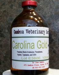
PHD Veterinary Service

PHD Veterinary Service

Contact Info

Dr. Porter @ 352-258-3571
portermi.dvm@gmail.com

Read more about Dr. Porter
And PHD Veterinary Services @


Showing posts with label horse veterinarian. Show all posts

Friday, January 31, 2014

Hip Subluxation in a Horse


A 10 year-old mare presented for sudden onset hind limb lameness. The mare was non-weight bearing on the right hind limb and within 24 hrs there was significant swelling centered over the right gluteal region. In Figure 1, the pelvis of a horse is pictured to identify the left (bottom blue arrow) and the right (top blue arrow) coxofemoral joints. In Figure 2 and 3, the pelvis has been rotated slightly to picture the area that is most commonly ultrasounded to assess the coxofemoral joint space. In Figure 3, the red line corresponds to the junction of the pelvis and the femor. The blue arrow corresponds to the coxofemoral joint space.

Figure 1

Figure 2


Figure 3
Figures 4 and 5 illustrate the views captured via ultrasound exam of the equine pelvis. This area is very difficult to adequately radiograph in a standing horse hence the ultrasound exam provides an opportunity to assess the coxofemoral joint space of a horse. As in Figure 3, the red line in Figure 5 corresponds to the juction of the pelvis and femur (coxofemoral joint space) which is also highlighted by the blue arrow. In the normal exam, the only bone structures commonly noted are the pelvis (long white line at bottom of view) and the greater trochanter of the femur (small white line at the top right of the image).
Figure 4

Figure 5
Figures 6 and 7 correspond to the right coxofemoral joint space of the lame horse. There is clearly and extra curved line in the image that corresponds to the "head" of the femur. Normally, the head of the femur is NOT visualized because it is within the acetabulum that makes up the coxofemoral joint. The ultrasound findings below are consistent with a subluxation of the coxofemoral joint space and very likely fracture of the acetabulum!  This injury carries a very poor prognosis for an adult horse; however, in miniature horses or very small ponies, surgical correction exists as an option with a guarded prognosis.

Figure 6


Figure 7


Friday, April 26, 2013

White Line Disease and Laminitis in a Horse!

Figure 1

 The radiograph in Figure 1 is that of a middle-aged gelding that presented for intermittent foot pain. One year prior, the gelding had developed a high fever of unknown origin (presumed viral). During the weeks that followed the high fever, the gelding was slow to walk in small circle and on concrete surfaces. Over several months, the gelding's lameness improved however he was consistently tender immediately after being trimmed by the farrier.  There are 2 disease processes identifiable in Figure 1. The first is chronic laminitis or founder.

Figure 2
 In Figure 2, the yellow lines correspond to the dorsal hoof wall and the dorsal aspect of the coffin bone. Normally, these lines are parallel to each other such that a horizontal line drawn across them would yield equal angles (angle A and angle B). However, when the coffin bone has "rotated" from its axis then the angles will differ as they do in Figure 2. Hence, the radiographic finding is consistent with laminitis (founder) and subsequent coffin bone rotation. This condition appears to be chronic, evidenced by the "flaring" of the tip of the coffin bone seen in the blue circle in Figure 3. This flaring often corresponds to small fractures of the tip of the coffin bone secondary to increase concussive forces over the toe region of the foot. The red arrow highlights the extremely thin sole which measured less than 5 mm!  Ideally, the sole depth at the tip of the coffin bone should measure between 10-15 mm in thickness.

Figure 3
 The second disease process noted in these radiographs is White Line Disease. This is evidence by the gas lucency traveling up the dorsal hoof wall (yellow arrows in Figure 4). The lateral radiograph is only 2 dimensional hence it is impossible to predict if this gas lucency is right down the middle of the hoof or if it wraps around the medial and lateral aspect of the hoof. Regardless, the gas lucency corresponds to separation of the hoof wall from the underlying tissue. White line disease may be the primary problem or secondary. In this case, it is likely secondary to the chronic founder/laminits however in primary cases, it can cause coffin bone rotation and result in laminitis. This condition results from the accumulation of dead/decaying material that slowly works its way from the sole surface up the hoof wall. The offending organism is believed to be in part anaerobic bacteria that thrive in a low-oxygen environment and produce gas that can be seen on a radiograph. Excessive gas accumulation may result in hoof wall separation from the coffin bone and coffin bone rotation.  Treatment involves aggressive removal (hoof wall resection) of ALL dead and decaying hoof wall to expose the offending organisms. Hoof wall resection along is very effective in treating this condition but it is typically paired with soaking the foot after resection in a variety of solutions to kill off the offending organisms.

Figure 4

Often, the early signs of white line disease can be detected at the time of hoof trimming and appear as a shallow crack at the toe region. The opposite foot of this horse had the very beginnings of white line disease evidenced by a small amount of gas lucency noted at the very tip of the hoof (Figure 5). When identified at this early stage, the condition is easily managed with regular balanced 4-pt trimmings by the farrier. Successful  management of white line disease is best accomplished through a farrier-veterinarian team effort. The farrier is the most important part of the team since they will be doing 99% of the work. The veterinarian will provide the radiographic studies to verify that adequate exposure of the diseased foot is accomplished.


Figure 5


Friday, January 4, 2013

Illegal Equine Supplements!!!!

Carolina Gold
Magnesium Sulfate
The supplements imaged above are both considered "performance enhancing" drugs and are banned from use in horses of all disciplines!! Why am I posting this information you ask? Please take a moment to read the story about "Humble" the pony that was recently published in The New York Times. This pony died suddenly at a major horse event after being administered an injectable "supplement" by the horse trainer. A published medication sheet indicated that the pony was treated with Estrone, Depo Provera, Banamine, and Dexamethasone multiple times in the 48 hrs preceding his sudden death! The medication that was administered just prior to the pony's sudden death was not listed and remains unknown. 


If this story does not upset you then read about all group of polo ponies that died due to an over-dose of a supplememt called Biodyl. This supplement contains vitamine B12, selenium, potassium, and magnesium. Oddly enough, this supplement is very popular in the illegal sport of rooster fighting!!


These stories are examples of the gross and inappropriate actions by horse trainers and veterinarians with regards to the type and frequency of medications given to show horses. Some supplements are "home made" such as Carolina Gold and there is no telling what substances are mixed into the bottle. Other supplements are FDA approved medications such as Magnesium Sulfate which are used in appropriately in horses to enhance performance. Too often, the result of excessive use is sudden death due to cardiovascular collapse.


I urge to know exactly what your horses are being treated with and ask the question "why". Please read below about the product called "Carolina Gold". Although this drug has not been associated with sudden death, it is illegal and its use may result in hazardous riding conditions!

The active ingredient in "Carolina Gold" is known as Gabapentin or "GABA".  When administered, it acts as an inhibitory neurotransmitter and therefore has the potential to be anxiolytic, analgesic, anticonvulsant, and sedative. In human medicine, GABA containing drugs are prescribed for epilepsy and neuropathic pain. Adverse side effects include dizziness  fatigue  and drowsiness, to name a few.

In veterinary medicine it has been used sparingly to control seizures in foals. In addition, it has been classified as a class 3 performance enhancing drug by the Association of Racing Commissioners International. Interestingly, this product was introduced into the performance horse world as a means to "calm" horses just before entering the show arena! In February of last year, the USEF officially banned this product from you in performance horses.

http://www.dressage-news.com/?p=14964


Thursday, December 27, 2012

Long toe/Under run heel!!

The image below is of a horse's foot that is suffering from a conformational problem known as "long toe-under run heels". This horse tends to grow significant amount of hoof wall within 4-5 weeks without growing normal heel. Although the heel regions grow, they tend to roll under the center of the hoof. As such, this horse and others like him are prone to heel pain and poor performance. In Figure 1 the center of rotation is marked by the blue line. Ideally, the length of foot that is in front of the blue line and behind should be equal. This would result in a horse that is balanced "cranial to caudal" with respect to the center of rotation. 


Figure 1

As the amount of foot behind  (B) or caudal to the center of rotation decreases compared to "A" there is  a significant increase in the amount of force that is applied across the heel regions. This results in the rolling under or "under run heels" that is imaged in Figure 1 and 2. When this horse is trimmed, the hoof wall needs to be trimmed back to the widest portion of the frog.  This may seem counter intuitive due to the apparent "lack of heel" however it is necessary to achieve "normal" heel growth. 
Figure 2

The radiograph below is that of horse that has heel pain and is lame when trotted in a small circle in both directions. The hoof tester exam noted a strong positive response across the bar region of both heels. Notice that the length of "B" is significantly shorter than "A". In order to correct this problem, corrective shoeing is a must!  The process begins with a balanced trim that includes lowering the heels to the widest portion of the frog. This is followed by applying a shoe that either has built-in break over such as a natural balance/equilibrium shoe or break over is increased manually be rolling the toe. In addition  the shoe is set extra full in an attempt to increase the length of "B" and hence support the caudal aspect of the foot. 
Figure 3

In Figure 4 the horse has been trimmed and re-set. Notice that the length of  "B" is nearly the same as "A". This horse is quite close to being balanced with regard to the center of rotation (blue line) and within 4-5 days returned to complete soundness. Finally, these horses that tend to have a long toes and under run heels need to be trimmed and re-set every 4-5 weeks. It is quite common that these horses are sound for the first 4 weeks after the farrier visit yet their performance begins to diminish as 5 to 6 weeks pass before they are trimmed and re-set. Although radiographs are not necessary to diagnose this problem, they are helpful for quantifying the extent of imbalance and documenting improvement after shoeing. 
Figure 4


Thursday, December 6, 2012

Displaced Soft Palate!!

Figure 1
The image above is an endoscopic pic of a middle-aged gelding that presented for a history of exercise intolerance. Apparently, during low level, forced exercise, the gelding would begin to make loud "gurgling" noises and become short of breath! In addition, the gelding would cough on a regular basis, especially when eating in the stall. 

Figure 2

 The image above is that from a normal horse. The yellow arrow is pointing to the epiglottis which is not visible in Figure 1 and Figure 3 due to the dorsal displacement of the soft palate. Normally, horses breathe through their nasal passages. The epiglottis helps keep the soft palate in position thus keeping the oral pharynx separate  from the nasal pharynx. However, when the soft palate becomes displaced and covers the epiglottis, the nasal pharynx and oral pharynx communicate directly. When this occurs, the horse suddenly begins to breath through its oral cavity which results in a gurgling sound and exercise intolerance.

Figure 3
The cause of the chronic dorsal displacement of the soft palate in this case is in part due to chronic inflammation of the soft palate (yellow arrow) and potentially an abnormal epiglottis (blue star). The epiglottis was not visualized in this exam and may require an oral endoscopy and skull radiographs to better determine if  there is a structural problem with the epiglottis. Management of this condition involves treating with systemic anti-inflammatory medications and medicated throat wash. If the soft palate remains displaced, surgical consultation is recommended. Yet another example of the benefit of an endoscopic exam for a horses that suffer from coughing, exercise intolerance, or nasal discharge.

Thursday, November 29, 2012

High Ring Bone!!

The radiographic image below is of the right hind pastern of a 4 year old mare that presented for a prepurchase exam. The mare had only been under saddle for 6 months. During the active exam, the mare cross-cantered when lunged to the left. In addition, the pastern area was "thicker" when compared to the opposite hind limb and the mare was moderately positive to flexion of the limb in question. Due to the suspicion of a significant problem, the right hind pastern was radiographed. The yellow arrows highlight new bone growth along the edges of the pastern joint. The bone growth or osteophytes are large and proliferative. These findings are indicative of advanced osteo-arthritis of the pastern joint otherwise known as "high ring bone". 


Figure 1
In Figure 2, the same osteophytes are noted as the course around the front of the pastern joint. The yellow arrows point to the osteophytes or irregular "white" structures along the edge of the pastern joint. 

Figure 2
Figure 3 and 4 are radiographs of a normal pastern joint. There are no osteophytes noted along the margins of the pastern joint. Notice in Figure 4 the smooth, curved border of the normal pastern joint. Interestingly, the pastern imaged in Figures 3 and 4 was radiographed because it palpated "thicker" than the opposite limb. Fortunately, there was no evidence of pastern arthritis. 

Figure 3

Figure 4
 In Figure 5 there is significant new bone growth or osteophyte development along the margins of the coffin joint. This is known as "low ring bone". Generally speaking, "high" or "low" ring bone is a significant finding during a prepurchase exam. Low ring bone is more common in draft breed horses. In addition, varying degrees of coffin joint arthritis, in active sport horses, is more common than pastern arthritis.

Figure 5
 High ring bone or pastern arthritis should be a concerning finding in any horse that is expected to carry a rider. Medical management of pastern osteoarthritis is limited to intra-articular treatment with cortisone or regenerative therapies. Unfortunately, the degeneration of the joint progresses rapidly and commonly results in chronic lameness. Surgical management involves fusion of the joint either with hardware or chemicals.