PHD Veterinary Service

PHD Veterinary Service

Contact Info

Dr. Porter @ 352-258-3571
portermi.dvm@gmail.com

Read more about Dr. Porter
And PHD Veterinary Services @


Saturday, August 9, 2014

Allergic Airway Disease in horses.

In the past 30 days (July 8 - August 8) I have examined 10 horses for the complaint of coughing and poor performance. The horses have ranged in age from 8 to 19 years of age. There has been no commonality with regards to sex or breed. However, they all live in Florida and it is the hottest month of the year!  Through a series of diagnostics which include a re-breathing exam, upper airway endoscopy, and trans-tracheal wash, all 10 horses have been diagnosed with allergic airway disease (AKA: heaves or COPD). None of these horses had previously been diagnosed with this condition. 
.
.
Upper trachea with large amounts of sputum present.

Lower trachea with thickened mucosa due to chronic inflammation

Allergic airway disease in horses typically results in coughing, increased respiratory effort, increased "abdominal" breathing, exercise intolerance and weight loss. If not managed properly, the condition worsens from year to year and can result in the death of the horse!! I have posted a blog previously discussing this disease and how it should be diagnosed and treated. Please click on the link below to read my blog from last year regarding this condition.

Allergic Airway Disease in a Horse


Friday, July 25, 2014

Collapsing Trachea in a Miniature Horse

A three year-old miniature stallion presented to PHD Veterinary Services for the complaint of open mouth breathing and a "honking" sound during respiration. At presentation, the stallion was agitated and was continuously pawing at his muzzle with his mouth open. The stallion was mildly sedated for the endoscopic exam and interestingly, his breathing became more normal. The most proximal trachea (Figure 1) appeared normal in diameter however the lumen quickly became narrowed (Figure 2). The trachea was nearly completely collapsed for approximately 6 cm at the level of the thoracic inlet. Distal to this area, the trachea appeared normal (Figure 3).

Figure 1

Figure 2

Figure 3
Normally, the lumen of the trachea is continuous, extending from the larynx to the primary bifurcation of the trachea (Figure 4). Unfortunately, the miniature horse breed is predisposed to the condition known as collapsing trachea. The condition is often diagnosed shortly after birth or can develop later in life if the horse is allowed to gain excessive weight. A common clinical complaint is that of a "honking" sound during heavy respiration. The severity of the collapsing trachea depends on the location and the extent of the narrowed lumen. The closer the area of collapse is to the chest cavity, the worse the prognosis. There are reported cases of collapsing tracheas in miniature horses that were repaired via intra- and extra-luminal stents. Unfortunately, they are short term fixes and often do not provide a long term solution.

Figure 4

Friday, July 11, 2014

Keratoma in a horse!!


A 10 year-old gelding presented to PHD veterinary services for a history of recurrent abscesses in the left front foot. Over a period of 6 months, the gelding developed 3 distinct abscesses which ruptured at the coronary band. At presentation the gelding was mildly lame and there was no active drainage from the most recent abscess rupture. A radiographic exam was performed to determine if there was a radiographic explanation for the development of multiple foot abscesses in the same foot.

Figure 1

The most notable finding in the radiographic exam was evidence of coffin bone rotation (Figure 1 and 2). In Figure 2, the red dotted lines should be parallel. The red line on the left corresponds to the dorsal hoof wall and the red line on the right corresponds to the dorsal aspect of the coffin bone. The reason they are not parallel is because the red line on the right has rotated in a down ward direction approximately 10-12 degrees. Evidence of coffin bone rotation suggests a history of laminitis or founder and this might explain the recurring foot abscesses. In addition, the yellow lines in Figure 1 highlight the hoof wall defect which developed secondary to the recurrent foot abscesses. There are thin areas that appear radiolucent (black lines) which extend from the dorsal hoof wall defect (yellow lines) down towards the bottom of the foot. These radiolucent lines may correspond to remnants of draining tracts from the recent abscesses.

Figure 2
Figure 3
In Figure 3, the radiographic beam is aimed downward through the hoof and coffin bone. The image on the left is the left foot and the image on the right is the right foot. The yellow dotted circles correspond to the tip of the coffin bone that appears to be more radiolucent (less bone) in the left foot compared to the right foot. This would suggest some sort of pathological process affecting the tip of the coffin bone in the left foot. The possibilities for these radiographic changes include laminitis, chronic abscess with bone infection (osteomyelitis), and keratoma. An MRI study of the foot was strongly recommended to determine the cause and provide key information for likely surgical exploration.

Figure 4
The gelding was referred to the University of Florida for MRI of the front feet. A large area of concern was highlighted in the MRI that was either a chronic abscess or a keratoma. Subsequently, a partial hoof wall resection was performed by Dr. Andrew Smith and his surgical team at University of Florida's college of veterinary medicine (Figure 4). Once the hoof wall was removed, a large keratoma (yellow dotted lines) was identified and removed. A keratoma consists of  a benign tumor of keratin or horn-producing cells. The keratoma will grow between the coffin bone and the hoof wall causing distortion of the hoof wall, recurrent foot abscesses, and laminitis. Surgical resection is the only option but should be preceded by an MRI to identify the keratoma and its dimensions. Prognosis is good for a full recovery assuming the entire keratoma is removed at the time of surgery.

Special thanks to the Dr. Andrew Smith and the University of Florida's radiology department!

Friday, June 20, 2014

Summer Sore Time is here!!

Although I have already written a blog concerning summer sores in horses (go to: http://michaelporterdvm.blogspot.com/2013/06/habronemiasis-summer-sore-in-horse.html) , the condition is a recurring one and it deserves another visit!! Below are several images from different horses that suffer from recurring habronemiasis or equine summer sores. The horse in Figure 1 suffers from recurring habronemiasis in all 4 limbs, whereas the horse in Figure 2 recently developed summer sores along his sheath!! These summers sores are medium in size and treatable with topical and systemic medication. However the horse in Figure 3 is suffering from a horrible summer sore of his lower limb that has not been treated but allowed to grow unabated and permanently disfigure the limb. It is frustrating to the horse owner that these lesions tend to return every year with the return of flies however if treated early and aggressively, management of this condition is quite do-able!!

Figure 1

Figure 2
For horses with recurring or first time habronemiasis, I recommend the following:

1: Systemic treatment with a dewormer containing Ivermectin. Treat twice, approximately 3-4 weeks appart.
2: Topical treatment with Dr. Porter's summer sore cream!!
3: If the summer sore is excessively large or in a location that is difficult to treat with topical medication, I recommend systemic therapy with corticosteroids. I prefer to medicate with a tapering dose of oral prednisolone over 30 days.

It is critical that the barn management incorporate a fly control program of their choosing. In addition, horses which suffer from recurring summer sores need to be treated the moment there is any redness or mild irritation present in the old summer sore sites!! If treated early, the lesion will typically respond favorably to topical medication without the need for systemic corticosteroids! 

Figure 3

Friday, June 13, 2014

Fractured Spinous Processes in a Horse

A 10 year-old gelding arrived to a horse show and was seriously injured during the unloading from the trailer. The rear gate was lowered and the horse remained standing in the trailer with the "butt bar" in place (Figure 1). For some unknown reason, the horse panicked and attempted to unload with the butt bar in place. The horse became trapped below the butt bar and struggled for several minutes before the owner was able to release the bar. Within moments of the event, the horse's withers became swollen and very sensitive to the touch. The gelding was treated with systemic anti-inflammatory agents and returned home in the trailer WITHOUT the  butt bar!


Figure 1
Two weeks after the injury, the gelding presented to PHD veterinary services for a radiographic evaluation of the withers. In Figure 2, the green line outlines the withers of the horse. The bone-like fingers projecting upward below the green line are the spinous processes which make up the withers. On physical exam there was minimal swelling over the withers however the horse was very sensitive to any pressure and there appeared to be a "dip" in the very bottom of the withers (yellow arrows).

Figure 2
Radiographic evaluation of the "dip" in the withers confirmed multiple fractures of 3 spinous processes (yellow arrows in Figure 3). The first spinous process that is fractured appears to be in several fragments and displaced from its normal position. The second spinous process appears fractured but not displaced and the third process appears to have an avulsion of the most proximal aspect of the spinous process. Although it is likely that these fractures will heal in time and be some what stable, the current concern is the development of a frustrating condition known as fistulated withers. This  occurs when a piece of fractured bone becomes devitalized of blood supply and subsequently "dies" becoming a sequestrum. The body's natural response is to rid itself of the sequestrum and will develop a draining wound that originates from the sequestrum. These wounds do NOT resolve until the sequestrum has been removed and this can be extremely challenging from a surgical stand point. Currently there is not evidence of a sequestrum however it will take several weeks/months to determine if one will develop. Prognosis for return to riding will depend the development of fistulated withers and how comfortable the horse is once the fractures have stabilized.

Figure 3

Friday, June 6, 2014

Rehabilitation of horses through controlled swimming!!

The images below were taken at The Sanctuary Sports Therapy and Rehabilitation Center in Ocala Florida. The horse that is entering the water and swimming across the equine swimming pool is in the process of being rehabilitated for a ligament injury. This horse has already under gone 8 weeks of strict stall confinement and several doses of extra-corporeal shockwave treatment for his injury. Horses that have spent extensive time in stall confinement are at risk of re-injury or a new injury once they are returned to work. This is because the are out of condition yet willing to run, buck, jump, and generally misbehave!! Unfortunately, most horses lack a good sense of self preservation!! 


 Depending on the type of injury, low-impact exercise is advantageous for rehabilitating horses after a lengthy confinement. Unfortunately, due to their significant weight and relatively small cross-sectional area of their hooves, horses tend to stress their musculoskeletal structure even at the walk. In addition, many can be fractious and unsafe to handle and/or ride after an extensive period of confinement. As such, swimming provides an excellent form of exercise that is very low-impact and safe. Fortunately, most horses enjoy swimming and are quick learners!!

My recommendation for horses returning from a lengthy period of stall confinement includes 30 days of swimming for 20 minutes, 4-6 days per week. In addition, the horse will be hand-walked for 10-20 minutes per day. The remainder of time the horse remains in stall confinement. In my opinion, this is far superior to simply turning the horse out in a small paddock for 30 days.


http://www.sanctuaryequinerehab.com/


Friday, May 23, 2014

Severe Cartilage Degeneration in a Horse

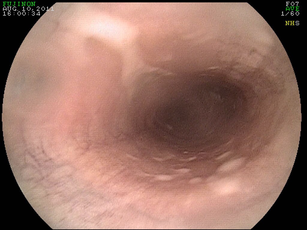
A 12 year-old show jumper mare presented to PHD veterinary services for the history of recurring forelimb lameness. In the past 2 years, the mare had the left front coffin joint treated with corticosteroids and hyaluronic acid every 6 months. However, the most recent treatment with intra-articular corticosteroids did not result in resolution of the lameness. On presentation, the mare was a grade 3/5 lame in the left front. The lameness was most evident when trotting on firm ground and in a circle to the right. The mare was NOT positive to hoof tester exam and was mildly positive to lower limb flexion. The coffin joint was "blocked" with carbocain and the mare's lameness improved by >85%. Radiographic exam of the left front foot only revealed evidence of mild osteoarthritis of the coffin joint.  (Figure 1).
Figure 1
Due to the following factors: 1) a history of poor response to intra-articular therapy, 2) a positive response to blocking the coffin joint, and 3) minimal data via the radiographic exam, it was recommended that the distal forelimb of this mare be evaluated via magnetic resonance imaging (MRI).  The MRI exam requires general anesthesia and generally costs between $1800-$2000; however the information gathered from an MRI exam is far superior than any other type of diagnostic exam! As such, I strongly recommend considering an MRI exam in cases that the CAUSE for the lameness has not been determined and a reasonable attempt has been made to treat and/or diagnose the problem. Figures 2-4 includes several images from the MRI exam involving this show jumper mare. A small, blue circle is included in each image to high light the large articular cartilage and subchondral bone defect that was discovered through the MRI study. In each image, the lesion appears as a small black circle at the surface of the bone. In addition to this finding, the MRI study noted additional evidence of chronic degeneration of the coffin joint. The full extent of the degeneration and the cartilage damage was NOT evident in the radiographic exam!!

Figure 2

Figure 3

Figure 4
Although the prognosis was poor for return to soundness, the mare was treated aggressively with various regenerative therapies and extended rest. Unfortunately, the mare remained significantly lame and it was determined that humane euthanasia was the best option due to the relentless foot pain. A post-mortom exam was performed and a very large area of abnormal cartilage loss (blue circle in Figure 5) was noted that corresponded to the MRI findings. This case represents yet another exam of the importance for getting an answer as to WHAT is causing the problem such that a prognosis can be provided followed by a proper course of action.

Figure 5
In the state of Florida, the most sensitive MRI for imaging in horses is ONLY available at the University of Florida's College of Veterinary Medicine. In addition, the board certified radiologists at the Veterinary College of Veterinary Medicine are especially well trained for the evaluation of MRI studies in horses! For more information about their services please go to:

http://largeanimal.vethospitals.ufl.edu/services/diagnostic-imaging/


Friday, May 16, 2014

Urinary Bladder tumor in a Horse

A 12 year old gelding presented to PHD veterinary services for the complaint of "dribbling" urine and intermittent bouts of urinating blood (hematuria). The horse had been treated with a combination of antibiotics and steroids which had resulted in resolution of blood in the urine however he continued to dribble urine. On presentation the physical exam was normal other than evidence of chronic urinary incontinence based on the accumulation of dried urine over the dorsum of both hind limb pasterns. A rectal exam was performed and a softball-size mass was palpated within the urinary bladder.

Figure1
Figure 2

Cystoscopic exam noted several small soft tissue masses leading into the urinary bladder with evidence of active, mild hemorrhage (Figure 1). Once the urinary bladder was entered with the scope, a mass was noted lying underneath the urine that was present in the urinary bladder (Figure 2). The urine was evacuated through the scope revealing a large, soft tissue mass that was firmly attached to the ventral or bottom aspect of the urinary bladder (Figure 3 and 4).

Figure 3

Figure 4
Urinary bladder neoplasia or cancer is rare in horses. The most common common type reported in horses is squamous cell carcinoma and it commonly associated with hematuria. Other types of cancer include transitional cell carcinoma, lymphosarcoma, leimyosarcoma and fibrous polyps. Management of urinary bladder neoplasia in horses involves surgical resection and possible intra-lesional chemotherapy; however the prognosis is poor for long term survival. This horse will be managed with systemic anti-inflammatory medications to reduce local inflammation within the urinary bladder and the proximal urethra.

Friday, May 9, 2014

Superficial Flexor Tendonitis in a Horse


A 12 year old mare presented for sudden onset forelimb lameness and swelling of the tendons in the right front limb. On palpation, the swollen tendons were significantly sensitive to palpation yet it was not possible to determine if it was the superficial flexor tendon (SDF) or the deep digital flexor (DDF) tendon. The mare was a grade 3/5 lame in the right front when trotted in a straight line on firm ground. Ultrasound exam of the soft tissue structures revealed a core-like lesion within the outside corner of the SDF tendon. In Figures 1-3, the core-like lesion appears as a black area (red circle) within the body of the SDF tendon (blue circle).  In Figure 2, the image to the right is a cross-sectional view of the tendon and the lesion is a small black circle within the body of the SDF tendon. In the image to the left in Figure 2, the tendon is in a longitudinal view and the lesion appears as a black streak through the body of the SDF tendon.This view is important because it highlights the over-all length of the lesion which in this case was 4-5cm long. The cross-sectional area measured in Figure 3 determined that the core-like lesion was approximately 20% of the entire cross-sectional area of the SDF tendon.

Figure 1

Figure 2

The mare was treated with ultrasound guided injection of Platelet rich plasma (PRP), stall rest, daily ice therapy and we will follow-up with 3 doses of extra-corporeal shock wave treatment. We anticipate a full return to work after the appropriate period of rehabilitation.

Friday, May 2, 2014

On-Line Pharmacy for PHD Veterinary Services!!


The above medications are just a few of the hundreds that my clients will be able to purchase via my on-line pharmacy!! The pharmacy will be open for business within the next 2 weeks and can be accessed at the following address:

PHD Veterinary Services On-Line Pharmacy

Once open for business, my clients will be able to register their pet's information and order the medications desired. I will receive an instant email to confirm authorization for the sale of the medications and the client will enter their credit card information. This pharmacy will contain hundreds of medications and nutraceuticals for cats, dogs, and horses! Once ordered the products will ship direct to my clients for quick delivery.

I anticipate that PHD Veterinary Service's on-line pharmacy will offer my clients an efficient and affordable method to purchase prescribed medications for their cats, dogs, and horses. There will be monthly sales on some of the more popular products so please visit my blog site to find out when the pharmacy is open for business!! Please visit the on-line pharmacy today and register your cats, dogs, and horses!