PHD Veterinary Service

PHD Veterinary Service

Contact Info

Dr. Porter @ 352-258-3571
portermi.dvm@gmail.com

Read more about Dr. Porter
And PHD Veterinary Services @


Friday, March 29, 2013

Coffin Bone De-Rotation!!

Approximately 6 weeks ago, a middle aged mare presented for chronic founder and non-weight bearing lameness. At presentation, there was significant rotation of the coffin bone in the lame foot (greater than 15 degrees) and reduction in the sole depth at the toe region (Figure 1-3). Due to the severe degree of coffin bone rotation and lameness it was determined that transection of the deep digital flexor tendon was the best option for the mare. The procedure was performed at the barn with sedation and a regional block.

Figure 1


Figure 2

Figure 3
In addition to transection of the deep digital flexor tendon, corrective shoeing was achieved by an expert farrier who specializes in foundered horses  (Sir Adam Whitehead). Corrective shoeing is as important if not MORE important than the surgical procedure and without it the procedure could not be a success! The mare's lameness has slowly improved over the past 4 weeks and follow up radiographs revealed complete DE-ROTATION of the coffin bone (Figure 4).  The palmar angle (blue line) is now approximately zero degrees with respect to the horizon and there has been a moderate increase in the sole depth at the toe region (yellow arrow).

Figure 4

In my experience, horses coffin bone rotation that exceeds 15 degrees benefit greatly from this procedure. Typically, these horses require 4-6 months from time of surgery before they are barefoot and sound in the pasture. This mare's initial recovery was slowed due to a sub-solar abscess that involved her entire sole and communicated with the coronary band. 

Friday, March 22, 2013

Guttural Pouch Fungal Infection Resolved!!

Back in January, a gelding presented for a history of recurrent nasal discharge. Endoscopic exam revealed the presence of a fungal infection within one of the guttural pouches (Figures 1 and 2) . A bacterial and fungal culture was performed on fluid/debris collected from the pouch. The bacterial culture was negative however the fungal culture was positive for Cladosporium spp. This species of fungi is very common in the environment, especially in the presence of moist or wet wood. In humans, it is a big player in fungi induced allergies. Interestingly, this species of fungi has not been described in the guttural pouch of a horse previously!

Figure 1

Figure 2
The gelding was treated with a specific anti-fungal medication given by mouth daily. After 4 weeks of treatment, there was a 50% reduction in the size of the fungal plaque and the degree of inflammation (Figures 3 and 4).


Figure 3

Figure 4
After 2 months of treatment there has been complete resolution of the fungal infection and inflammation (Figures 5 and 6). The stylohyoid bone remains slightly thickened compared to the opposite stylohyoid bone (Figure 7) however this will like resolve over the next 3-6 months. This case is unusual in several ways. First, the species of fungi has not been described before in the guttural pouch of a horse, secondly, most guttural pouch fungal infections include bleeding (epistaxis) thirdly, there are few reports of successful treatment of guttural pouch fungal infections in horses with JUST systemic anti-fungals.

Figure 5

Figure 6

Figure 7

Thursday, March 14, 2013

Fibrosarcoma in a horse!


A 14 year old gelding presented for a complaint of recent lameness and swelling of the right knee or carpus. There was no history of trauma but a plumb size swelling was palpated along the outside of the knee. The gelding resented direct pressure over the knee and any manipulation of the knee. When radiographed, there were no significant abnormalities noted with regards to the bones or articular surfaces that make up the carpus. However, a moderate soft tissue swelling was noted along the dorsal (front) and lateral (outside) aspect of the carpus (Blue circles in Figures 3-4). 

Figure 1

Figure 2

Figure 3

Figure 4
An ultrasound exam was performed on the soft tissue swelling and a hypoechoic (dark) soft tissue structure was identified (Figure 5). This structure appeared well demarcated and was consistent with a mass or tumor which was below the surface of the skin yet outside the joint capsule of the carpus.

Figure 5

When the ultrasound image is flipped into the same projection as the radiograph, the soft tissue mass aligns perfectly with the soft tissue swelling noted on the digital radiograph (Figure 6). The red line corresponds to the middle carpal joint and helps demonstrate how close the tumor is to the joint yet does NOT communicate with the joint.

Figure 6
A core biopsy was taken of the soft tissue mass through a small skin incision directly over the area in question and submitted for analysis.  The histopathology report indicated that the core biopsy was consistent with a fibrosarcoma. This type of tumor is not common in horses and although it does not commonly spread to other regions of the horse, it can be locally destructive and aggressive. As such, it was determined that the immediate course of action was intra-lesional injections of a potent chemotherapy drug. Due to the close proximity with the joint capsule, surgery was considered too risky at this point. The gelding has been treated 1x with a chemotherapy medication and a follow-up exam is expected within 2-3 weeks. To be continued...

Thursday, March 7, 2013

Osteochondroma in a horse!!

 The radiograph in Figure 1 corresponds to the carpus of the horse which became lame ONLY after heavy work. Once he was lame, the lameness was resolved by injecting carbocaine into carpal joints. Careful radiographic examination of the carpus and the surrounding structures noted a bony protuberance along the distal, palmar aspect of the radius (Figure 1 and 2). This finding is consistent with an osteochondroma formation. In humane medicine, an osteochondroma is defined as " an abnormal, solitary, benign growth of bone and cartilage, typically at the end of a long bone". In horses, osteochondroma formation is not common however when it does occur, the occurs commonly along the lower end (distal) aspect of the radius. In this location, the osteochondroma may cause irritation to the surrounding soft tissue structures including the carpal sheath. 
Figure 1

Figure 2
In Figure 3 and 4, an ultrasound exam was performed of the distal radius to determine if the osteochondroma was the source of the lameness. Two irregular lines (yellow lines) can be seen on the ultrasound exam which are consistent with the bony protuberances noted in the radiographs. In addition, the surrounding tissue is irregular with pockets of edema and there is a large accumulation of fluid within the carpal sheath (Red star). 

Figure 3


Figure 4
When the left and right forelimbs were compared via ultrasound (Figure 5), there is no evidence of an osteochondroma in the normal leg (right) compared to the left leg. A needle was placed into the pocket of fluid within the carpal sheath and blood tinged synovial fluid was collected. Following fluid aspiration, the carpal sheath was treated with cortisone, antibiotic, and Hylartin V. Most osteochondromas in horses are surgically removed and the prognosis is good for full return to work.

Friday, March 1, 2013

Equine Herpes Virus-1


What is EHV-1?

The acronym EHV-1 refers to Equine Herpes Virus -1 which is one of 4 varieties of the equine herpes virus complex (EHV-1, EHV-2, EHV-3, and EHV-4). EHV-4 is associated with upper respiratory disease in horses where as EHV-1 is associated with respiratory, neurologic, abortion, and foal death. EHV-3 is also known as coital exanthema and is a sexually transmitted disease in horses. This family of viruses is found in horses all over the world and it is unclear why some horses develop the neurologic form of this disease complex.

How is EHV-1 transmitted?

Transmission of the virus from one horse to another is dependent on 1: direct contact (nose to nose), 2: indirect contact via contaminated items and 3: aerosolized fluids (coughing or sneezing). Aerosolized fluids may travel up to 35 feet! The virus may survive for up to 30 days in the environment if the conditions are ideal. Once horses are infected they become latent carriers for the remainder of their life. They may become spontaneous "shedders" during periods of stress!

What are the clinical signs?

Incubation period is typically 6-8 days (time from exposure to onset of clinical signs) however it has been reported to be as long as 21 days!

Common clinical signs may include fever, depression, inappetance, upper respiratory infection, and abortion.
Neurologic signs range from temporary ataxia (in-coordination), urinary incontinence, rear limb weakness (dog sitting), complete paralysis and death. Death may occur within 24 hours of the onset of neurologic signs!!


How do you diagnose and treat horses with EHV-1?

Detection of EHV-1 in horses may be through PCR testing of nasal swab or blood, serologic testing, virus isolation and post-mortem exam.

Treatment is based supportive care which may include IV fluid therapy, anti-inflammatory medication and in some cases anti-viral drugs. There is no specific medication to treat EHV-1 in horses!!

Does vaccination protect horses from EHV-1?

There is no commercially available vaccine that prevents the disease! However there are several vaccines which are believed to reduce nasal shedding and hence limit the spread of disease. These include Rhinoimmune (Boehringer Ingelhein), Calvenza (BI), Pneumorabort-K (Pfizer) and Prodigy (Merck). Vaccination during an outbreak is recommended ONLY if there is a history of being vaccinated previously with these vaccines. Recommended to vaccinate every 3-6 months.

What should you do in the face of an outbreak?


Encourage barn personnel to disinfect clothing, shoes, and hand-wear at the entry and exit of all barn areas.

Monitor rectal temperature daily in horses exposed to known EHV-1 positive horses.

If your horse has been exposed to a horse known to be positive for EHV-1, a 21 day isolation protocol is necessary! Isolation area must consider the potential for a 35 foot range of aerosolized mucus.


Additional information may be viewed at the following sites:

AAEP and EHV-1

UF Veterinary Hospital and EHV-1

Department of Agriculture in Florida and EHV-1

Thursday, February 21, 2013

Laminitis in a horse!!

 The following radiographs are from a gelding that has been lame for nearly 1 year. The gelding has been treated for laminitis/founder and despite efforts by the farrier and veterinarian, the gelding's conditions has worsened. He is non-weight bearing on one forelimb at the time of my exam. The right front foot is imaged in Figure 1 and the left front foot in Figure 2. In Figures 3 and 4 the palmar angle (angle "A") is measured in both front feet. The palmar angle corresponds to the angle that the bottom or palmar surface of the coffin bone makes with the horizon. In both front feet, the palmar angle measures approximate +23-25 degrees. Normally, the palmar angle may range between zero to +5 degrees. Abnormal palmar angles may be negative or greater than +7-8 degrees. However, even though the right and left front feet share the same palmar angle, it is for two very different reasons!

Figure 1 (Right front)

Figure 2 (Left front)
Figure 3 (Right front)
Figure 4 (Right front)
The most likely causes for an increased palmar angle include coffin bone rotation and coffin joint contracture. The difference between these two conditions is key to making the correct diagnosis. In the case of laminitis, the coffin bone rotates under the influence of the deep digital flexor tendon and due to a lack of attachment between the coffin bone and the lamina (i.e. laminitis). In Figure 5, the degree of coffin bone "rotation" is estimated based on the difference between angles "A" and "B". Normally, the hoof wall (red line over "A")  is parallel with the coffin bone (red line over "B"). When these two lines are parallel, the angles equal each other and there is zero degrees of coffin bone rotation. Therefore, the problem with the left front limb in Figure 5 is laminitis and secondary coffin bone rotation.

Figure 5 (Left front)
The right front foot has zero degrees of coffin bone rotation however the palmar angle is the same as in the left front foot. This due to coffin joint contracture and is the definition of a "Club foot". In Figure 6, the lines corresponding to the hoof wall and coffin bone are parallel and as such angle  "A" equals angle "B". Figure 7 diagrams the concept of coffin joint contracture that results in an angle of contracture (angle "A"). Typically, a club foot or coffin joint contracture does not result in non-weight bearing lameness but rather a life long commitment to proper shoeing and trimming. In this case, first glance of the radiographs might be deceiving because both front feet have an abnormal palmar angle, but it is the left front foot with the coffin bone rotation and life threatening laminitis! The deep digital flexor tendon was transected in the left front limb and follow-up radiographs will follow!
To be continued......

Figure 6 (Right front)
Figure 7 (Right front)


Friday, February 15, 2013

Equine Hock Arthritis

Figure 1
The radiograph above (Figure 1) is of a horse's hock or tarsus. This view is also known as the "Lateral" view or side view. The equine hock joint is made up of 4 individual joints which are labelled above. The bottom two joints are also known as the distal hock joints and include the distal inter-tarsal joint (DIT) and the tarsal metatarsal joint (TMT). When "injecting" hocks, these are the most common joints treated and are often referred to as the "upper and lower hock joints". Degenerative joint disease (DJD) or osteo-arthritis is most common in the distal hock joints and  often results in poor performance plus/minus lameness. Bog spavin is the term that refers to increased joint fluid within the tibiotarsal joint. This is the "high motion" joint of the hock and is NOT commonly injected with "hock injections". However if increased fluid is noted, a radiographic exam is definitely indicated prior to instituting a treatment plan. Generally speaking, arthritis of the distal hock joints is more acceptable and can be managed with intra-articular therapies. However, arthritis of the tibiotarsal joint and/or the proximal inter-tarsal joint is more concerning and is reason for concern when predicting future performance.

Figure 2

In Figure 2 and 3, there are radiographic changes that indicate osteo-arthritis of the tarsal metatarsal joint space. These changes were noted during a prepurchase exam of a 5 year old horse. The horse was sound during the exam and did NOT respond to hock/stifle flexion. The million dollar question is what to recommend to the buyer based on these findings. In my clinical experience, most horses with these changes will eventually need intervention via intra-articular cortisone injections. I believe that horses with these changes are MORE likely to need hock injections than those with "normal" radiographs. However, these findings are not necessarily a negative prognostic indicator with regards to the horse's future performance. 

With management, i.e, hock injections, these horses can compete at the highest level and succeed!  As such, these findings do NOT constitute a FAILING grade during the prepurchase exam however the buyer needs to be prepared for the strong likelihood of routine "maintenance". This can easily result in hundreds to thousands of dollars per year that should be considered into the price of the horse. 

Figure 3

Thursday, February 7, 2013

Proximal Suspensory Desmitis in a Horse

The ultrasound images below are from a teenage gelding that presented for a 3 month history of mild, forelimb lameness. The lameness would improve with rest but would return soon after the gelding was returned to work. On physical exam, the proximal suspensory palpated sensitive, just below the carpus (knee) of the right forelimb. The gelding was not lame in a straight line however when lunged in a circle to the left, a mild lameness (2/5) was noted in the right forelimb. The lameness improved approximately 50% after blocking the lower limb however when the proximal suspensory ligament was blocked, the gelding was sound. Ultrasound exam of the tendons and ligaments revealed a focal area of decreased echogenicity (dark spot) which was consistent with inflammation and edema within the proximal suspensory ligament. 

Figure 1

Figure 2
In Figure 1, there is a cross sectional image of the superficial flexor tendon (SDF), deep digital flexor tendon (DDF), distal check ligament, and the proximal suspensory ligament. This image was made approximately 10cm below the knee or carpus. In Figure 2, the same area is imaged in cross section and in longitudinal plane. The same lesion (dark spot) can be seen in both images which is consistent with a "real" lesion versus an artifact. In the longitudinal view (right image in Figure 2), the origin of the suspensory is highlighted by the blue arrows and the edema is noted by the dark fibers just above the blue arrows. In Figure 3, the cross sectional image is slightly obliqued to visualize the inside or medial aspect of the limb. The lesion within the suspensory ligament is more apparent in this image and is represented by the dark blue circle within the yellow circle (suspensory ligament) in Figure 3.


Figure 3

Figure 4

The history and lameness exam findings are "classic" for a forelimb proximal suspensory ligament injury. Often the lameness is most noticeable when the affected limb is on the outside of the circle and the lameness will improve temporarily with rest. The prognosis for this injury is "good" however will require rest, ice therapy, corrective shoeing and a specific rehabilatory program. Adjunct therapies include shockwave treatment and platelet rich plasma (PRP) injections.


Friday, February 1, 2013

Gastric Ulcers in a horse

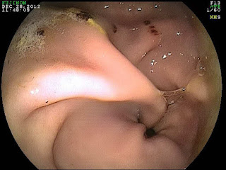
The endoscopic images below are from a teenage gelding that presented for a complaint of aggressive  behavior while grooming. The gelding was not displaying any classic signs of colic nor was there a decline in food intake or a report of weight loss. When the stomach was examined, several bleeding ulcers were noted surrounding the pyloric sphincter. This region of the stomach includes the passage from the stomach into the small intestine. The gelding was treated with 45 days of Gastrogard and returned to normal behavior.


 The important message from this case is that gastric ulcers in horses can present like most anything! Classically, they present as low grade colic associated with feeding plus or minus weight loss. However, in my experience clinical signs associated with gastric ulcers have included poor performance, dull hair coat, excessive water intake, sour behavior under saddle, aggressive behavior while being groomed, colic, parking out, frequent posturing to urinate, teeth grinding, and weight loss.


I strongly recommend a gastric exam which includes visualization of the pyloris to determine if gastric ulcers are the source of the clinical complaint. This involves 24 hrs of fasting and a trained clinician with a 3 meter gastroscope. If ulcers are discovered, the only treatment proved to be effective is Gastrogard medication for at least 30 days! I typical recommend 30 days of a full dose followed by 2 weeks of a half dose.


Friday, January 25, 2013

Guttural Pouch Mycosis in a horse!!

The endoscopic images in Figure 1 and 2 are that of a normal guttural pouch in a horse. There are 2 guttural pouches in the horse and their role is not clearly defined. However, there are several very important structures which course through the guttural pouches. These include large veins and arteries plus critical cranial nerves (Figure 2) . Each guttural pouch is divided into a medial and lateral compartment by a unique bone named the stylohyoid bone. This bone articulates with the base of the skull, just below the ear drum and is part of the support structure for the tongue and larynx! As such, when the horse moves its tongue the articulation between the stylohyoid bone and the base of the skull moves as well. The large blood vessels located within the guttural pouch are important for bringing oxygenated blood to the brain and draining deoxygenated blood from the brain. Equally important are the cranial nerves that course through the guttural pouches. These nerves are essential for a proper swallowing reflex, sensation to the face, and balance.

Figure 1
Figure 2
The endoscopic images in Figures 3-5 are from a gelding that presented for a history of purulent nasal discharge that responded to antibiotic treatment. The endoscopic exam was requested as a follow-up to make sure there was nothing lurking in the horse's upper airway. When the right guttural pouch was entered, a large white plaque was noted covering the entire stylohyoid bone. There was minimal discharge within the pouch and there appeared to be mold covering the surface of the plaque!!

Figure 3
Figure 4
In Figures 4 and 5, the proximity of the fungal plaque with the large blood vessels and important nerves can be seen. Normally, fungi seek out vascular tissue and slowly erode the walls of vessels which can result in low grade bleeding and if it is a large artery, sudden death!! Commonly, horses with fungal infection or mycosis of the guttural pouch present with a history of a bloody nose (epistaxis), however this horse did not. When the plaque was disturbed with the scope, the underlying tissue was exposed and the inflammation was evident.  A bacterial and fungal culture was performed on the fluid recovered during the guttural pouch lavage and a fungi was recovered yet the identity is still being worked out. This condition is difficult to treat and is currently being managed with systemic antifungals and guttural pouch lavage. Stay tuned!!!!

Figure 5