PHD Veterinary Service

PHD Veterinary Service

Contact Info

Dr. Porter @ 352-258-3571
portermi.dvm@gmail.com

Read more about Dr. Porter
And PHD Veterinary Services @


Showing posts with label suspensory ligament. Show all posts

Friday, September 27, 2013

Hind Limb Proximal Suspensory Ligament Desmitis in a Horse

A 15 year-old mare presented for a 3 week history of rear-limb lameness that was associated with a "drop" of the rear fetlock joint. On presentation there was moderate swelling of the lower limb, just below the hock joint and the mare was lame at the walk. In addition, there was a 90 degree drop of the fetlock/pastern axis as noted in Figure 1.

Figure 1

The primary mechanism involved in "suspending" the fetlock joint and maintaining the proper fetlock/pastern axis is the suspensory ligament (Figure 2). The suspensory ligament originates just below the hock (red arrow) and initially is one structure (body) that travels down the back of the lower limb (yellow arrow). Approximately half way down the canon bone the suspensory ligament splits into a medial (inside) and lateral (outside) branch. The suspensory branches attach to the sesamoid bones which are located just behind and below the fetlock joint. As such, the suspensory ligament helps "suspend" the fetlock joint and a  proper fetlock/pastern axis.

Figure 2
An ultrasound exam was performed to evaluate the entire suspensory ligament. The origin or proximal suspensory ligament is imaged in cross-section in Figures 3-6. The proximal suspensory ligament of the affected limb is grossly enlarged (yellow circle) and the fiber pattern is a mixed pattern with significant edema and evidence of active inflammation! There is a black and grey swirl pattern noted in the proximal suspensory ligament (tissue inside the yellow circle) of the affected limb which is indicative of severe changes.


Figure 3


Figure 4

 When compared to the normal limb, the significant increase in the size of the proximal suspensory ligament is evident. In this case the affected suspensory ligament was 2x the "normal" size. These ultrasound findings confirm the diagnosis of proximal suspensory desmitis of the hind limb. The prognosis for this injury is poor for return to riding and guarded for return to pasture soundness. Once the fetlock has "dropped" the physical changes to the suspensory ligament CANNOT be reversed!! Treatment is aimed at slowing the progress of the condition and attempting to provide pain relief to the horse. In my experience, corrective shoeing is the MOST important aspect of managing this condition.

Figure 5
  
Figure 6
A "fish tail" bar shoe is strongly recommended for this condition. The shoe should be set extra full such that approximately 1.5 to 2 inches of shoe extended BEHIND the heel bulb. Any kind of a wedge is CONTRAINDICATED in this condition! In addition, daily treatment with ice packs over the proximal suspensory ligament followed by topical Surpass cream are indicated to reduce inflammation and provide pain relief. With corrective shoeing, adequate pain relief, and supervised turn-out, these horses may return to pasture soundness however such a condition carries a guarded prognosis.

Figure 7


Friday, October 26, 2012

Not All Splints Are Created Equal!!


The radiographs below are from the forelimbs of a lame horse. The horse had prominent "bumps" on the inside of both canon bones and only one was sensitive to direct pressure (right front). These bumps were consistent with osseous or bony callous formation over the splint bones which are also know as "splints". The  splints had been present for over 45 days and despite moderate rest, the horse remained lame. Specifically, the horse was lame in the right front limb when trotted in a circle to the left! Careful exam of these "splints" revealed that in addition to being painful to pressure over the callous, the horse was positive to pressure over the suspensory ligament at the level of the fractured splint.


Ultrasound exam of the soft tissue structures near the fractured splint revealed an inflamed suspensory ligament. The image below highlights the suspensory ligament (yellow arrow). The areas of black are consistent with edema/fluid accumulation within the ligament and are adjacent to the bony callous. In addition  to the presence of edema/fluid, the fiber pattern is irregular. These findings are consistent with moderate inflammation of the suspensory ligament, most likely due to physical interference via the bony callous. 


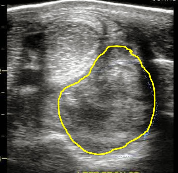
The ultrasound image below compares the left and right suspensory ligaments at the level of the corresponding "splints". A clear difference can be seen between the suspensory ligaments (area under the yellow curves) of the lame leg (RF) versus the non-lame leg.


The horse was scheduled for surgery and the offending splint bone was removed. At the time of surgery, moderate inflammation of the suspensory ligament was confirmed. The gelding underwent an extensive period of rehabilitation consisting of rest and controlled exercise. Three months post surgery the gelding is back to work and completely sound. Special thanks to Dr. Tim Lynch at Peterson and Smith Equine Hospital for his surgical expertise and great collaborative effort!

This case is not unique and reminds us that we should pay close attention to ANY "splint" development. Most often, splints resolve without the development of lameness or significant complications however if the appropriate care is not considered, the likelihood of complications does increase. Appropriate care includes forced rest, ice therapy, topical Surpass, and shockwave treatment.

Thursday, May 3, 2012

Suspensory Break Down!

The suspensory ligament has been significantly stretched and strained, resulting in the "dropped fetlock" conformation seen below. The suspensory ligament (blue structure in drawing below) originates just below the hock in the hind limbs and the carpus in the forelimbs. The ligament splits into a medial (inside) and lateral (outside) suspensory branch. The split occurs approximately half way down the metatarsus. Each suspensory branch inserts on a sesamoid bone at the level of the fetlock and play a role in "suspending" the fetlock. For the fetlock to drop, the suspensory branch and body have been strained and maybe even ruptured. For definitive diagnosis, an ultrasound exam is performed that will evaluate the entire suspensory ligament. If the process is slow (months to years), it may result in reduced performance and low grade lameness. However, if sudden, significant lameness is associated with the limb or limbs affected. Unfortunately, this is a progressive condition that will ultimately result in retirement of the horse and may be life-threatening. Management of this condition includes reduced exercise, daily ice therapy, corrective shoeing, and daily pain management. This condition carries a poor prognosis for return to work and a guarded prognosis for over-all soundness.


The blue structure depicts the suspensory ligament in the forelimb. Note the upper region is one structure (body) which then splits into two branches.


These are known as "fish tail" shoes and are quite effective in reducing the strain on the suspensory ligaments. For obvious reasons, this type of shoe, with significant heel extension, can only be used in the hind limbs.