PHD Veterinary Service

PHD Veterinary Service

Contact Info

Dr. Porter @ 352-258-3571
portermi.dvm@gmail.com

Read more about Dr. Porter
And PHD Veterinary Services @


Showing posts with label tendon ultrasound. Show all posts

Friday, April 11, 2014

Superficial Flexor Tendonitis in a Horse

A middle-aged gelding presented to PHD veterinary services for tendonitis of the superficial flexor tendon (SDF). The tendonitis was a result of an tendon-sheath penetrating injury and was associated with a severe tendon sheath infection. The tendon sheath infection was treated aggressively and effectively by an equine surgeon. An ultrasound exam was performed along the plantar aspect of the hind fetlock and a large, core-like lesion was identified (Figure 1, black area within red circle). The lesion consisted of disrupted tendon fiber and edema. Several options were considered to help the "healing process" involving the SDF tendonitis. These included extra corporeal shock wave treatment, platelet rich plasma (PRP) injection, stem cell injection, and/or concentrated bone marrow injection. The decision was made to treat the lesion by injecting a mixture of concentrated bone marrow mixed with PRP product. The bone marrow was collected from the horse's sternum and concentrated on site (Figure2). In addtion, a blood sample was collected from the horse and PRP was harvested on site. The PRP was mixed with the concentrated bone marrow and then injected into the lesion via ultra-sound guided technique.

Figure 1

Figure 2


In addition to the PRP+bone marrow injection, the horse was fitted with a corrective shoe that provided significant heel extension (Figure 3 and 4). This type of shoe is also known as a "fish tail" show and will reduce the "load" across the fetlock joint by supporting the lower limb. Horses recovering from tendon sheath infections and SDF tendonitis tend to avoid fully loading the back of the foot which may result in lower limb contracture. The fish tail shoe allows the horse to fully load the foot in a more comfortable fashion. The horse was walked daily and otherwise kept in stall confinement. In addition, the leg was treated daily with cold compresses.

Figure 3

Figure 4
Approximately 30 days after the PRP+bone marrow injection, the horse's limb was re-ultrasounded. There has been significant "filling in" of the lesion (Figure 5). Although the area that was the lesion (red circle) remains identifiable, there has been a significant improvement in tendon fiber alignment and the edema (black) has nearly completely resolved! Although this represents a tremendous amount of improvement in just 30 days, there remains a significant period of rehabilitation before the ultimate out come of this case can be reported.  This case provides an example of a regenerative therapy that combines PRP and progenitor stem cells (bone marrow) for the treatment of tendonitis! The advantages of this approach versus stem cell treatment include reduced cost, on site harvesting and same-day treatment.


Figure 5


Friday, September 27, 2013

Hind Limb Proximal Suspensory Ligament Desmitis in a Horse

A 15 year-old mare presented for a 3 week history of rear-limb lameness that was associated with a "drop" of the rear fetlock joint. On presentation there was moderate swelling of the lower limb, just below the hock joint and the mare was lame at the walk. In addition, there was a 90 degree drop of the fetlock/pastern axis as noted in Figure 1.

Figure 1

The primary mechanism involved in "suspending" the fetlock joint and maintaining the proper fetlock/pastern axis is the suspensory ligament (Figure 2). The suspensory ligament originates just below the hock (red arrow) and initially is one structure (body) that travels down the back of the lower limb (yellow arrow). Approximately half way down the canon bone the suspensory ligament splits into a medial (inside) and lateral (outside) branch. The suspensory branches attach to the sesamoid bones which are located just behind and below the fetlock joint. As such, the suspensory ligament helps "suspend" the fetlock joint and a  proper fetlock/pastern axis.

Figure 2
An ultrasound exam was performed to evaluate the entire suspensory ligament. The origin or proximal suspensory ligament is imaged in cross-section in Figures 3-6. The proximal suspensory ligament of the affected limb is grossly enlarged (yellow circle) and the fiber pattern is a mixed pattern with significant edema and evidence of active inflammation! There is a black and grey swirl pattern noted in the proximal suspensory ligament (tissue inside the yellow circle) of the affected limb which is indicative of severe changes.


Figure 3


Figure 4

 When compared to the normal limb, the significant increase in the size of the proximal suspensory ligament is evident. In this case the affected suspensory ligament was 2x the "normal" size. These ultrasound findings confirm the diagnosis of proximal suspensory desmitis of the hind limb. The prognosis for this injury is poor for return to riding and guarded for return to pasture soundness. Once the fetlock has "dropped" the physical changes to the suspensory ligament CANNOT be reversed!! Treatment is aimed at slowing the progress of the condition and attempting to provide pain relief to the horse. In my experience, corrective shoeing is the MOST important aspect of managing this condition.

Figure 5
  
Figure 6
A "fish tail" bar shoe is strongly recommended for this condition. The shoe should be set extra full such that approximately 1.5 to 2 inches of shoe extended BEHIND the heel bulb. Any kind of a wedge is CONTRAINDICATED in this condition! In addition, daily treatment with ice packs over the proximal suspensory ligament followed by topical Surpass cream are indicated to reduce inflammation and provide pain relief. With corrective shoeing, adequate pain relief, and supervised turn-out, these horses may return to pasture soundness however such a condition carries a guarded prognosis.

Figure 7


Friday, June 7, 2013

Superficial digital Flexor Tendonitis (Bowed tendon) in a Horse!

A  twenty year-old mare presented for sudden, severe swelling of the left front lower limb. The mare was nearly non-weight bearing lame and had been seen galloping around her paddock moments before. Prior to my exam, the mare was stalled for 7 days and treated with ice and systemic non-steroidal mediation (phenylbutazone). On presentation there was a noticeable "profile" to the lower portion of the forelimb (Red line:Figure 1). The mare was moderately positive to pressure over this swelling and was lame at the walk. On palpation, there was no distinction between the superficial digital flexor tendon and the deep digital flexor tendon.
Figure 1
Ultrasound examination of the lower limb, beginning just below the knee (carpus) revealed inflammation of the superficial digital flexor tendon (SDF). The inflammation was moderate just below the knee (Figure 2) however became severe further down the leg (Figure 5). In Figures 2 and 3, moderate to severe edema (dark tissue) is noted with pockets of disrupted fiber pattern. The image in Figure 4 is of the opposite limb for comparison to /the abnormal limb. In Figure 5, the SDF tendon (Red arrows) is severely enlarged and there is no apparent pattern with regards to the fiber pattern. These findings are consistent with a near rupture of the SDF tendon.

Figure 2

Figure 3

Figure 4

Figure 5
Although the outward and inward findings are severe, the prognosis for pasture soundness is good. It will take 6-12 months for this type of injury to heal with a fibrotic scar but the mare is likely to return to soundness as a pasture horse. Unfortunately, her prognosis as a riding horse is poor and it is not recommended to return this mare to any type of forced work due to her age and degree of injury. Tendonitis of the superficial flexor tendon can occur in various regions of the lower limb. The closer to the knee/carpus (High bow), the worst the prognosis, even for pasture soundness. This is due to the constant irritation of the tendon when the horse advances the limb. In addition, horses that suffer a "bowed" SDF tendon are at increased risk for another injury to the same tendon and it commonly occurs above or below the original injury. My recommendation for rehabilitative care includes the following:

1: Limited activity: preferred stall rest with hand walking for 2 months followed by small paddock turn-out
2: Daily ice therapy for 2-3 weeks
3: Topical non-steroidal cream: Surpass
4: Consider Extra-corporeal shockwave treatment and PRP injection.

Friday, May 31, 2013

Distal Check Ligament Injury in a Horse

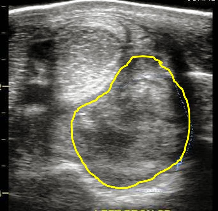
A 14 year-old, warm-blood mare presented for a sudden swelling along the upper region (2 inches below the carpus on the back of the leg) of the flexor tendons in the left forelimb. The mare was mildly to moderately positive to palpation of the swelling yet no obvious lameness was observed when trotted in hand. Careful palpation of the area isolated the swelling to the distal check ligament/deep digital flexor tendon area of the proximal metacarpus. Approximately 2-3 inches below the knee/carpus of a horse, 4 soft tissue structures can be identified on ultrasound exam (Figure 1). The ligaments and tendons are typically assessed in a cross-sectional plane (Figure 1-3) and in a longitudinal plane (Figure 4). The most superficial is the superficial digital flexor tendon (SDF), followed by the deep digital flexor tendon (DDF), followed by the distal check ligament, and finally the deepest soft tissue structure which is the proximal suspensory ligament. The first two structures (DDF and SDF) are tendons and should appear identical with regards to brightness (echogenicity) on ultrasound. The deep structures (check ligament and suspensory) are ligaments and tend to be brighter on ultrasound exam when compared to the tendons. The overall echogenicity should be the same or homogeneous throughout the body of the tendons and ligaments.

Figure 1
In Figure 2, a clear, black lesion is identified along the outside/lateral aspect of the distal check ligament. The entire image appears slightly side-ways or oblique in order to optimize visualization of the ligament injury (Red circle in Figure 3). The "black hole" or core lesion corresponds to an area of ligament fiber disruption, edema, and possible blood accumulation.

Figure 2
Figure 3

In Figure 4, the same area is examined in a longitudinal plane to assess the extent of the fiber disruption. The area of disrupted ligament appears to include nearly the entire length of the distal check ligament. Although there is significant edema and contrast between the check ligament and the surrounding structures, the ligament appears to be intact. On a scale of mild, moderate, and severe, I would grade this as moderate with regards to degree of injury to the distal check ligament.

Figure 4
The image in Figure 5 is a simultaneous cross sectional and longitudinal image of the injured ligament.  Of all the soft tissue structures to injure in this region of the horse's limb, the distal check ligament is the least important with regards to long term prognosis and return to work. In my experience, many horses with this injury demonstrate little if any lameness at the time of injury but an obvious swelling is always present. Lameness in these horses is typically reserved for the severe injuries which involve a tear of the distal check ligament.
Figure 5
Care for horses with this type of injury include forced rest, ice, topical anti-inflammatory medication (Surpass cream), platelet rich plasma injection (PRP), and extra-corporeal shockwave treatment. At a minimum, the horse needs to be stalled with hand walking only for 6-8 weeks followed by 6-8 weeks of a controlled rehab program. During this time, daily ice treatment is ideal. However, I have had patients that did not agree to the stall rest and were merely turned out for several months. These horses healed with a prominent thickness to the ligament but were sound and return to full work. In my experience, the combination of PRP injection followed by a series of shockwave treatment results in a smaller scar (thickening of the ligament). I have been involved in 2 cases that resulted in surgery to remove the distal check ligament. In one case, the ligament had been injected with cortisone which resulted in  abscessation of the ligament and in the second case, the injury was not allowed to heal properly before returning the horse to work!  Although these types of injury have a good prognosis for full return to work, the horse does need time off and I strongly recommend regular evaluations for soundness and follow-up ultrasound exam.