PHD Veterinary Service

PHD Veterinary Service

Contact Info

Dr. Porter @ 352-258-3571
portermi.dvm@gmail.com

Read more about Dr. Porter
And PHD Veterinary Services @


Friday, January 25, 2013

Guttural Pouch Mycosis in a horse!!

The endoscopic images in Figure 1 and 2 are that of a normal guttural pouch in a horse. There are 2 guttural pouches in the horse and their role is not clearly defined. However, there are several very important structures which course through the guttural pouches. These include large veins and arteries plus critical cranial nerves (Figure 2) . Each guttural pouch is divided into a medial and lateral compartment by a unique bone named the stylohyoid bone. This bone articulates with the base of the skull, just below the ear drum and is part of the support structure for the tongue and larynx! As such, when the horse moves its tongue the articulation between the stylohyoid bone and the base of the skull moves as well. The large blood vessels located within the guttural pouch are important for bringing oxygenated blood to the brain and draining deoxygenated blood from the brain. Equally important are the cranial nerves that course through the guttural pouches. These nerves are essential for a proper swallowing reflex, sensation to the face, and balance.

Figure 1
Figure 2
The endoscopic images in Figures 3-5 are from a gelding that presented for a history of purulent nasal discharge that responded to antibiotic treatment. The endoscopic exam was requested as a follow-up to make sure there was nothing lurking in the horse's upper airway. When the right guttural pouch was entered, a large white plaque was noted covering the entire stylohyoid bone. There was minimal discharge within the pouch and there appeared to be mold covering the surface of the plaque!!

Figure 3
Figure 4
In Figures 4 and 5, the proximity of the fungal plaque with the large blood vessels and important nerves can be seen. Normally, fungi seek out vascular tissue and slowly erode the walls of vessels which can result in low grade bleeding and if it is a large artery, sudden death!! Commonly, horses with fungal infection or mycosis of the guttural pouch present with a history of a bloody nose (epistaxis), however this horse did not. When the plaque was disturbed with the scope, the underlying tissue was exposed and the inflammation was evident.  A bacterial and fungal culture was performed on the fluid recovered during the guttural pouch lavage and a fungi was recovered yet the identity is still being worked out. This condition is difficult to treat and is currently being managed with systemic antifungals and guttural pouch lavage. Stay tuned!!!!

Figure 5


Thursday, January 17, 2013

Dysphagia and Aspiration Pneumonia in a horse!!


The endoscopy images below were from a young gelding that presented for a history of coughing while eating. In addition, an intermittent nasal discharge was present and the gelding was described as a "hard keeper". In Figure 1, a small amount of feed material was noted along the floor of the soft palate, just in front of the epiglottis. In addition, feed particles were noted on the epiglottis and the walls of the pharynx. These findings alone are not 100% indicative of a dysphagic (difficulty swallowing) horse however, generally speaking there should NOT be any feed material in the pharynx or larynx. 

Figure 1
When the endoscope was passed through the larynx into the trachea (wind pipe) copious amounts of feed material mixed with mucus was noted on the walls of the trachea. In Figure 2, green and brown feed material  can be seen adhered to the wall of the trachea. As the endoscope was pass further down the trachea, feed material mixed with mucus remained present in significant amounts. 

Figure 2
At the very end or deepest region of this image (Figure 3) is the primary bifurcation of the trachea which occurs just in front of the heart. The presence of feed material at this level of the airway is consistent with aspiration and a trans-tracheal wash was performed to confirm pneumonia.  

Figure 3

Dysphagia and aspiration pneumonia are quite uncommon in horses and may be secondary to a neuropathy affecting the nerves that control the swallow reflex. The most common disease processes in horses that may affect these nerves include equine protozoal myeloencephalitis (EPM), cervical spine trauma, trauma to the guttural pouches, temporohyoid osteopathy (THO), strangles, encephalitis, and basisphenoid bone fracture. Unfortunately, the prognosis is poor for horses with this condition. The likelihood that they will recover their normal swallow reflex, regardless of the cause, is low!  In addition, the aspiration pneumonia that results from the dysphagia  is a recurring problem that typically results in their failure to thrive!


Friday, January 11, 2013

Cervical Spine Fracture

The radiographic images below are from a young gelding that was found by the owner walking "strangely" in the pasture. There was a large swelling present along the neck region, just behind the right ear and the gelding was having difficulty raising his head above the horizon. There was no knowledge of recent trauma. When examined, the gelding WAS walking and the "strange" gait was better described as moderate ataxia in all  limbs. The gelding was able to elevate his head with assistance but there was minimal lateral movement of this cervical spine. 


The radiographic exam revealed a catastrophic fracture of cervical spine #2 (C2), also known as the axis. The fracture line (yellow line) appears to travel directly through the spinous process of  C2 and through the body of C2. Centered in the body of the cervical spine is the vertebral canal which houses the spinal cord. Typically, an injury to this region of the spine results in trauma to the spinal cord and varying degrees of neurologic deficits. 

Due to the severity of the fracture in this case, one might have predicted that the trauma to the spinal cord would have resulted in severe neurologic deficits such as complete recumbency or even sudden death! Although the gelding was still standing, there was significant instability of the cervical spine and the prognosis for life was poor, hence humane euthanasia was elected. 


Very sad case!!


Friday, January 4, 2013

Illegal Equine Supplements!!!!

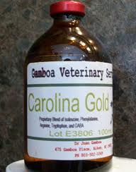
Carolina Gold
Magnesium Sulfate
The supplements imaged above are both considered "performance enhancing" drugs and are banned from use in horses of all disciplines!! Why am I posting this information you ask? Please take a moment to read the story about "Humble" the pony that was recently published in The New York Times. This pony died suddenly at a major horse event after being administered an injectable "supplement" by the horse trainer. A published medication sheet indicated that the pony was treated with Estrone, Depo Provera, Banamine, and Dexamethasone multiple times in the 48 hrs preceding his sudden death! The medication that was administered just prior to the pony's sudden death was not listed and remains unknown. 


If this story does not upset you then read about all group of polo ponies that died due to an over-dose of a supplememt called Biodyl. This supplement contains vitamine B12, selenium, potassium, and magnesium. Oddly enough, this supplement is very popular in the illegal sport of rooster fighting!!


These stories are examples of the gross and inappropriate actions by horse trainers and veterinarians with regards to the type and frequency of medications given to show horses. Some supplements are "home made" such as Carolina Gold and there is no telling what substances are mixed into the bottle. Other supplements are FDA approved medications such as Magnesium Sulfate which are used in appropriately in horses to enhance performance. Too often, the result of excessive use is sudden death due to cardiovascular collapse.


I urge to know exactly what your horses are being treated with and ask the question "why". Please read below about the product called "Carolina Gold". Although this drug has not been associated with sudden death, it is illegal and its use may result in hazardous riding conditions!

The active ingredient in "Carolina Gold" is known as Gabapentin or "GABA".  When administered, it acts as an inhibitory neurotransmitter and therefore has the potential to be anxiolytic, analgesic, anticonvulsant, and sedative. In human medicine, GABA containing drugs are prescribed for epilepsy and neuropathic pain. Adverse side effects include dizziness  fatigue  and drowsiness, to name a few.

In veterinary medicine it has been used sparingly to control seizures in foals. In addition, it has been classified as a class 3 performance enhancing drug by the Association of Racing Commissioners International. Interestingly, this product was introduced into the performance horse world as a means to "calm" horses just before entering the show arena! In February of last year, the USEF officially banned this product from you in performance horses.

http://www.dressage-news.com/?p=14964


Thursday, December 27, 2012

Long toe/Under run heel!!

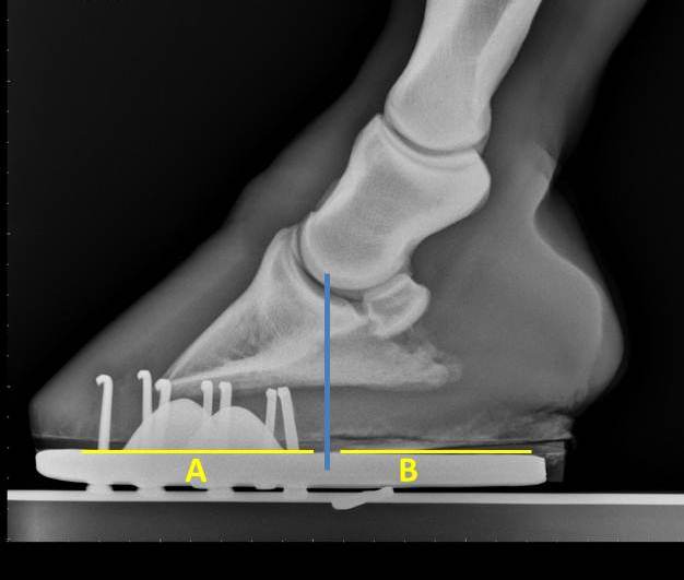
The image below is of a horse's foot that is suffering from a conformational problem known as "long toe-under run heels". This horse tends to grow significant amount of hoof wall within 4-5 weeks without growing normal heel. Although the heel regions grow, they tend to roll under the center of the hoof. As such, this horse and others like him are prone to heel pain and poor performance. In Figure 1 the center of rotation is marked by the blue line. Ideally, the length of foot that is in front of the blue line and behind should be equal. This would result in a horse that is balanced "cranial to caudal" with respect to the center of rotation. 


Figure 1

As the amount of foot behind  (B) or caudal to the center of rotation decreases compared to "A" there is  a significant increase in the amount of force that is applied across the heel regions. This results in the rolling under or "under run heels" that is imaged in Figure 1 and 2. When this horse is trimmed, the hoof wall needs to be trimmed back to the widest portion of the frog.  This may seem counter intuitive due to the apparent "lack of heel" however it is necessary to achieve "normal" heel growth. 
Figure 2

The radiograph below is that of horse that has heel pain and is lame when trotted in a small circle in both directions. The hoof tester exam noted a strong positive response across the bar region of both heels. Notice that the length of "B" is significantly shorter than "A". In order to correct this problem, corrective shoeing is a must!  The process begins with a balanced trim that includes lowering the heels to the widest portion of the frog. This is followed by applying a shoe that either has built-in break over such as a natural balance/equilibrium shoe or break over is increased manually be rolling the toe. In addition  the shoe is set extra full in an attempt to increase the length of "B" and hence support the caudal aspect of the foot. 
Figure 3

In Figure 4 the horse has been trimmed and re-set. Notice that the length of  "B" is nearly the same as "A". This horse is quite close to being balanced with regard to the center of rotation (blue line) and within 4-5 days returned to complete soundness. Finally, these horses that tend to have a long toes and under run heels need to be trimmed and re-set every 4-5 weeks. It is quite common that these horses are sound for the first 4 weeks after the farrier visit yet their performance begins to diminish as 5 to 6 weeks pass before they are trimmed and re-set. Although radiographs are not necessary to diagnose this problem, they are helpful for quantifying the extent of imbalance and documenting improvement after shoeing. 
Figure 4


Thursday, December 13, 2012

Round Two!! Pharyngeal Phythiosis

Figure 1
The endoscopic image in Figure 1 is from a middle aged quarter horse that presented for abnormal noise during exercise. The entire pharynx is obstructed by multiple granulomas.  Initial biopsy results were consistent with pythiosis and the gelding was treated with systemic anti-fungal medication for several months. 

Figure 2
Recheck endoscopy after 45 days revealed a significant improvement (Figure 2) ; however, treatment was discontinued prematurely and a final endoscopy was not performed.

Figure 3

Over 1 year after the initial presentation, the abnormal noise returned along with bloody nasal discharge. The pale yellow nodules have increased in size and number. Multiple small yellow granuoles are noted through out the pharynx and there is evidence of mild bleeding. The horse will again be treated with systemic anti-fungal medication. A follow up exam to follow!!

Thursday, December 6, 2012

Displaced Soft Palate!!

Figure 1
The image above is an endoscopic pic of a middle-aged gelding that presented for a history of exercise intolerance. Apparently, during low level, forced exercise, the gelding would begin to make loud "gurgling" noises and become short of breath! In addition, the gelding would cough on a regular basis, especially when eating in the stall. 

Figure 2

 The image above is that from a normal horse. The yellow arrow is pointing to the epiglottis which is not visible in Figure 1 and Figure 3 due to the dorsal displacement of the soft palate. Normally, horses breathe through their nasal passages. The epiglottis helps keep the soft palate in position thus keeping the oral pharynx separate  from the nasal pharynx. However, when the soft palate becomes displaced and covers the epiglottis, the nasal pharynx and oral pharynx communicate directly. When this occurs, the horse suddenly begins to breath through its oral cavity which results in a gurgling sound and exercise intolerance.

Figure 3
The cause of the chronic dorsal displacement of the soft palate in this case is in part due to chronic inflammation of the soft palate (yellow arrow) and potentially an abnormal epiglottis (blue star). The epiglottis was not visualized in this exam and may require an oral endoscopy and skull radiographs to better determine if  there is a structural problem with the epiglottis. Management of this condition involves treating with systemic anti-inflammatory medications and medicated throat wash. If the soft palate remains displaced, surgical consultation is recommended. Yet another example of the benefit of an endoscopic exam for a horses that suffer from coughing, exercise intolerance, or nasal discharge.

Thursday, November 29, 2012

High Ring Bone!!

The radiographic image below is of the right hind pastern of a 4 year old mare that presented for a prepurchase exam. The mare had only been under saddle for 6 months. During the active exam, the mare cross-cantered when lunged to the left. In addition, the pastern area was "thicker" when compared to the opposite hind limb and the mare was moderately positive to flexion of the limb in question. Due to the suspicion of a significant problem, the right hind pastern was radiographed. The yellow arrows highlight new bone growth along the edges of the pastern joint. The bone growth or osteophytes are large and proliferative. These findings are indicative of advanced osteo-arthritis of the pastern joint otherwise known as "high ring bone". 


Figure 1
In Figure 2, the same osteophytes are noted as the course around the front of the pastern joint. The yellow arrows point to the osteophytes or irregular "white" structures along the edge of the pastern joint. 

Figure 2
Figure 3 and 4 are radiographs of a normal pastern joint. There are no osteophytes noted along the margins of the pastern joint. Notice in Figure 4 the smooth, curved border of the normal pastern joint. Interestingly, the pastern imaged in Figures 3 and 4 was radiographed because it palpated "thicker" than the opposite limb. Fortunately, there was no evidence of pastern arthritis. 

Figure 3

Figure 4
 In Figure 5 there is significant new bone growth or osteophyte development along the margins of the coffin joint. This is known as "low ring bone". Generally speaking, "high" or "low" ring bone is a significant finding during a prepurchase exam. Low ring bone is more common in draft breed horses. In addition, varying degrees of coffin joint arthritis, in active sport horses, is more common than pastern arthritis.

Figure 5
 High ring bone or pastern arthritis should be a concerning finding in any horse that is expected to carry a rider. Medical management of pastern osteoarthritis is limited to intra-articular treatment with cortisone or regenerative therapies. Unfortunately, the degeneration of the joint progresses rapidly and commonly results in chronic lameness. Surgical management involves fusion of the joint either with hardware or chemicals.

Friday, November 16, 2012

Pythiosis!

The endoscopic images below are of the pharynx of a horse with a 30 day history of a bloody nose. The nasal discharge consisted of bright red blood mixed with some purulent debris. The middle-aged gelding lives in a pasture that includes multiple low-lying areas that have been flooded with stagnant water. In addition, the gelding has suffered from failure to thrive most likely due to a poor appetite.


Figure 1
The endoscopic exam revealed bleeding tissue that surrounded the pharynx but was most severe along the dorsal wall of the pharynx. There was purulent debris mixed with the bleeding tissue and multiple small, white granuoles were noted within the bleeding tissue (Figure 2) .  Possible disease processes for this condition include fungal granuloma, cancer, bacterial abscess, and pythiosis.  Pythiosis is caused by the invasion of tissues by the pathogen pythiosis insidiosum and may affect external and internal tissues. The pathogen typically gains access to the horse via exposure to stagnant water that contains p. insidiosum.  As such the lower limbs (pasterns, coronary bands, and fetlocks) are most commonly affected. Once infected, the wound becomes increasingly inflamed with persistent bloody discharge. In addition, the lesions are very puritic resulting in additional inflammation via the horse biting and scratching at the wounds. Within the large areas of inflammation are firm, yellow granuoles also known as "kunkers".  This condition can be very deadly and requires aggressive treatment. For external cases, surgical removal and topical treatment are common. In addition a vaccine is available that has proven effective for cases of equine pythiosis.

Figure 2
However, pythiosis of the pharynx is not treatable with surgical resection or the common topical medications. Fortunately, systemic antifungals have proven effective if treated early and for the appropriate amount of time. The endoscopic images below are from the same horse approximately 4 weeks into the systemic anti-fungal treatment protocol. I recommend a specific protocol that involves  6 weeks of a tapering dose and a follow up endoscopic exam. In the past 8 years I have treated nearly a dozen cases of pharyngeal pythiosis. In every case there was a body of stagnant water available to the horses and all but 1 case responded to the systemic anti-fungal medication. For more information regarding the condition and the vaccine check out the following link: http://pythium.pavlab.com/subpage5.html


Figure 3

Figure 4


Friday, November 9, 2012

Phenylbutazone Toxicity!!

The yearling pictured below was being treated with Phenylbutazone for a severe lameness. The yearling weighed approximately 750 lbs and was being treated with 2 grams of Phenylbutazone twice per day for 2 weeks! Although the lameness improved, the yearling stopped eating and developed mild colic symptoms, severe oral ulceration (Figure 1) and eventually generalized edema. 

Figure 1

The serum levels of total protein and albumin were well below normal values and an abdominal ultrasound revealed severe edema of the right dorsal colon (Figure 2) . These findings were consistent with an advanced case of right dorsal colitis secondary to excessive phenylbutazone administration. Most often, when we discuss "ulcers" in horses we are referring to gastric or stomach ulcers. These types of ulcers may be caused by drugs such as banamine and phenylbutazone or are often a result of poor nutritional management. However, colonic ulcers may also be caused by such drugs, especially phenylbutazone. These types of ulcers are poorly understood and often difficult to diagnose in a standing horse. A typical history is that of a horse that presents for mild, recurrent colic, mild to severe diarrhea, ill-thrift, and a history of recent phenylbutazone treatment. Although the dose of phenylbutazone in this case was excessive, some horses appear to be super sensitive to the drug and may develop right dorsal colitis on an accepted dose of phenylbutazone. 

Figure 2
 Sadly, the yearling described could not be saved and was euthanized. A necropsy was performed and severe ulceration of the right dorsal colon (Figure 3 and 4) was discovered along with the marked edema of the colon wall (Figure 5). These large button-like ulcers cannot be detected via an ultrasound exam and would require surgical exploration of the colon to diagnose. The edema of the right dorsal colon wall can be diagnosed via trans-abdominal ultrasound and is secondary to the significant loss of serum protein. The protein is lost through the GI tract due to the inflamed colonic surface.
Figure 3

Figure 4

Figure 5
 Phenylbutazone toxicity can range from mild to severe. Mild cases are rather common and may present as only mild colic symptoms following administration of the drug. As such, careful observation should be made at all times when treating with these types of drugs and a reduction in dose and frequency of administration should be considered sooner than later! Horses suffering from colonic ulcers do NOT respond to gastric ulcer medication such as ranitidine and omeprazole. There is no specific medication for this condition. Treatment includes termination of phenylbutazone treatment, de-bulking the diet (more pellets and less hay), and time. Prognosis is guarded for advanced cases and good for mild cases.